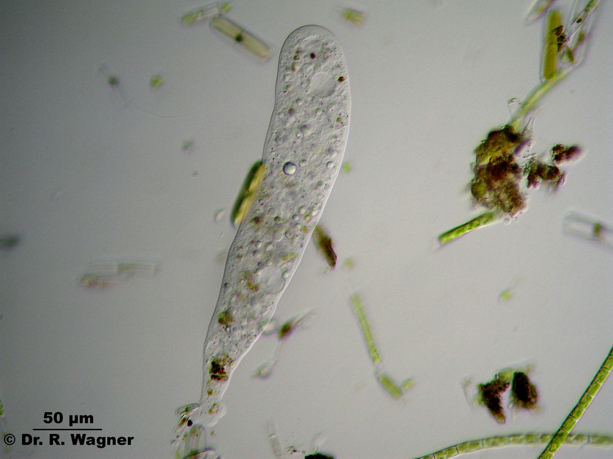
Especially several contractile vacuoles can be seen
Amoeba, (video, WMV-format, 2.9 MB) botanical garden university Duesseldorf, March 2008
changes in shape are shown here
an amoeba, probably amoeba hylobata national horticultural show Duesseldorf, April 2006
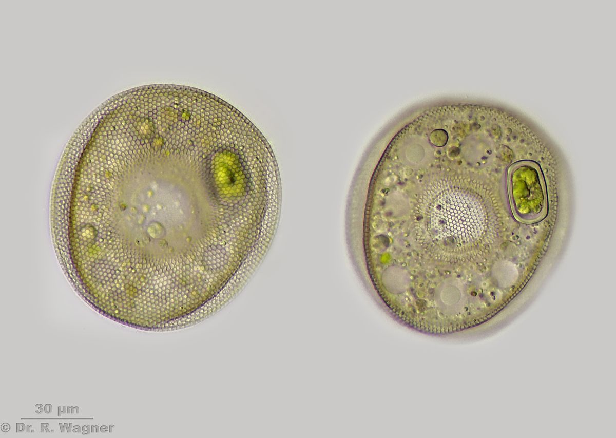
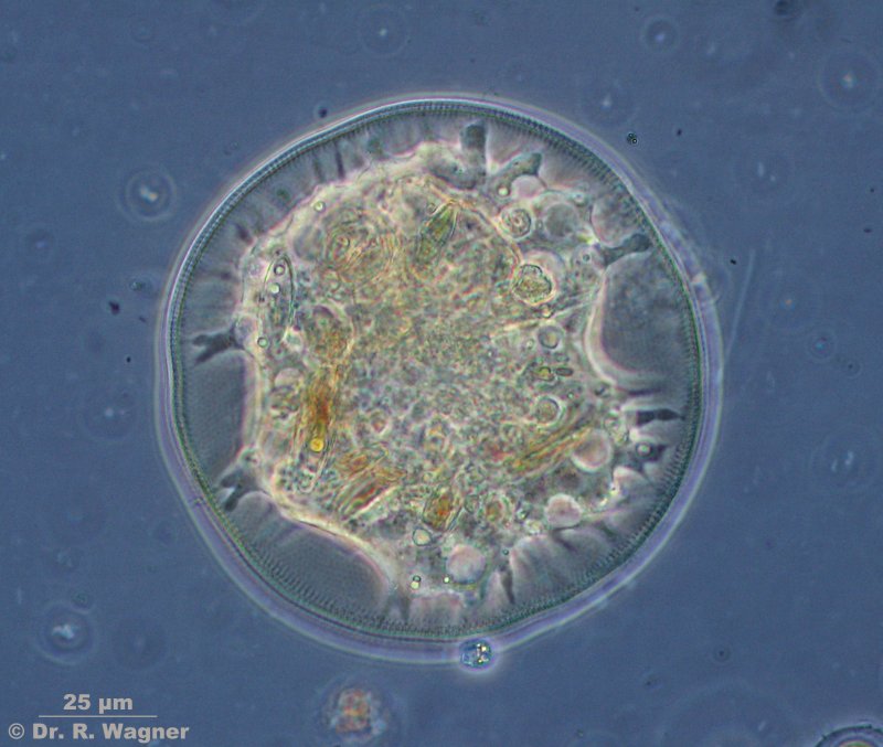
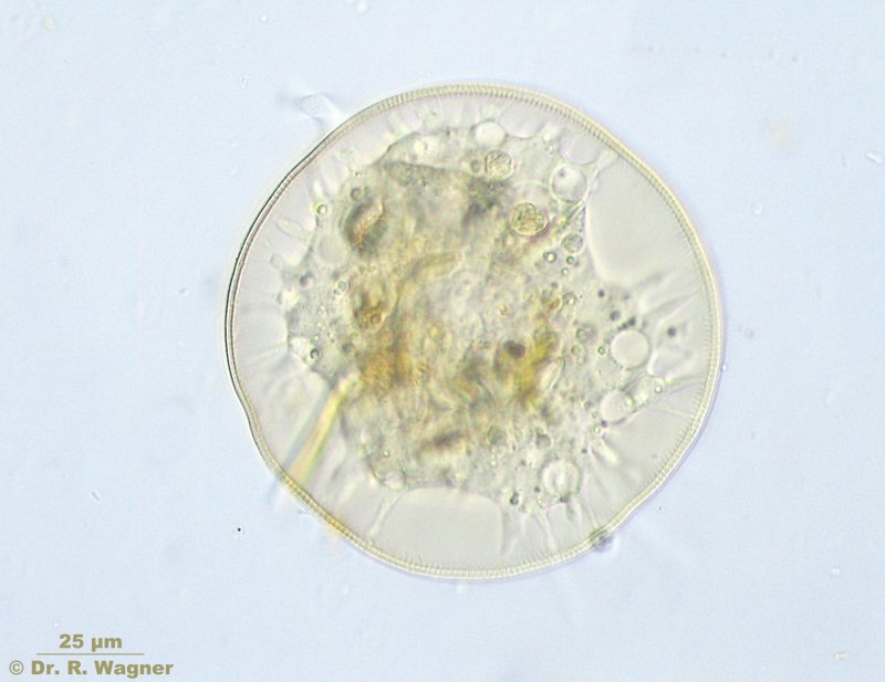
arcella spec. gallows-moor, September 2005
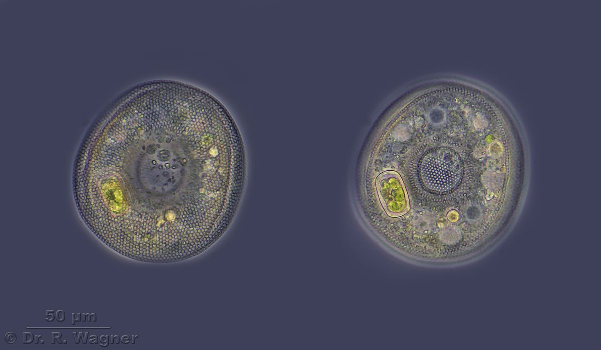
arcella gibbosa national horticultural show Duesseldorf, January 2007
bottom view, amoeba itself in its lorica
arcella gibbosa national horticultural show Duesseldorf, January 2007
side view with 2 pseudopodia
arcella gibbosa national horticultural show Duesseldorf, January 2007
Pseudopodia in phase contrast.
arcella gibbosa national horticultural show Duesseldorf, January 2007
Movement of the arcella, divx coded video (2,9 MB). First the colorless ectoplasma
moves in front of the pseodopodia, followed by the grained entoplasma.
Arcella spec. heathland pond near Roermond, May 2015
Arcella spec. heathland pond near Roermond, May 2015
Phasecontrast
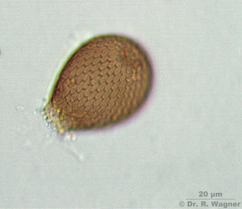
Assulina muscorum Garden pond, between mosses, February 2008
Phase contrast
Assulina muscorum Garden pond, between mosses, February 2008
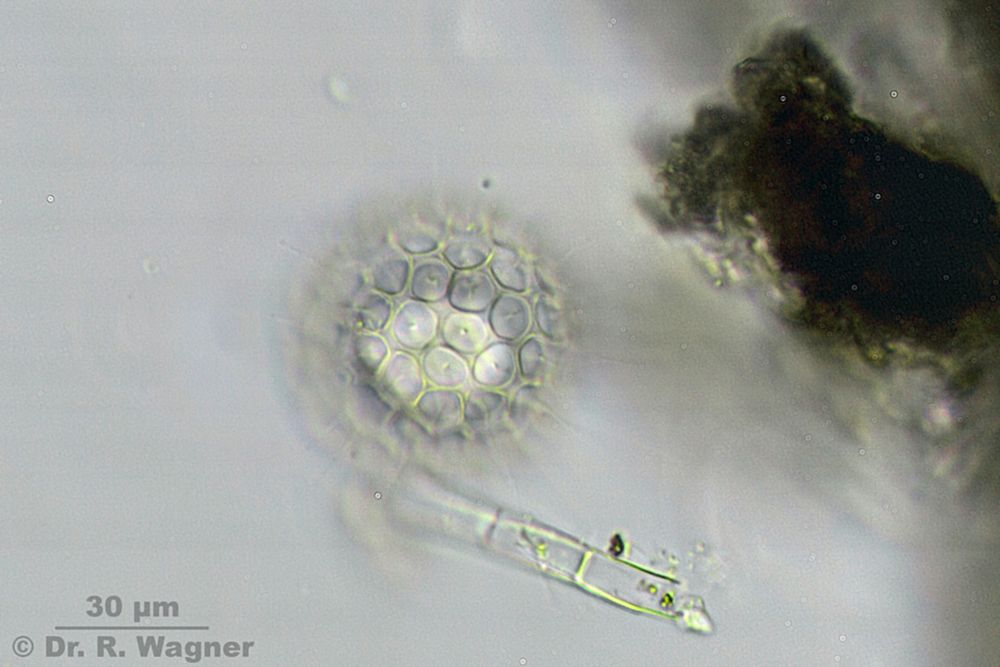
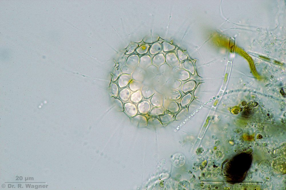
Clathrulina elegans Gardenpond August 2016
Focus on the cagestructure
Clathrulina elegans Gardenpond August 2016
Focus on the cagestructure
Clathrulina elegans Gardenpond August 2016
Focus on the center
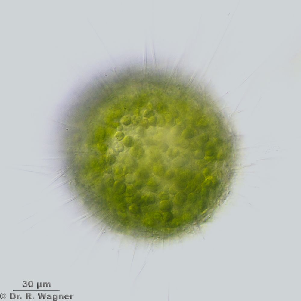
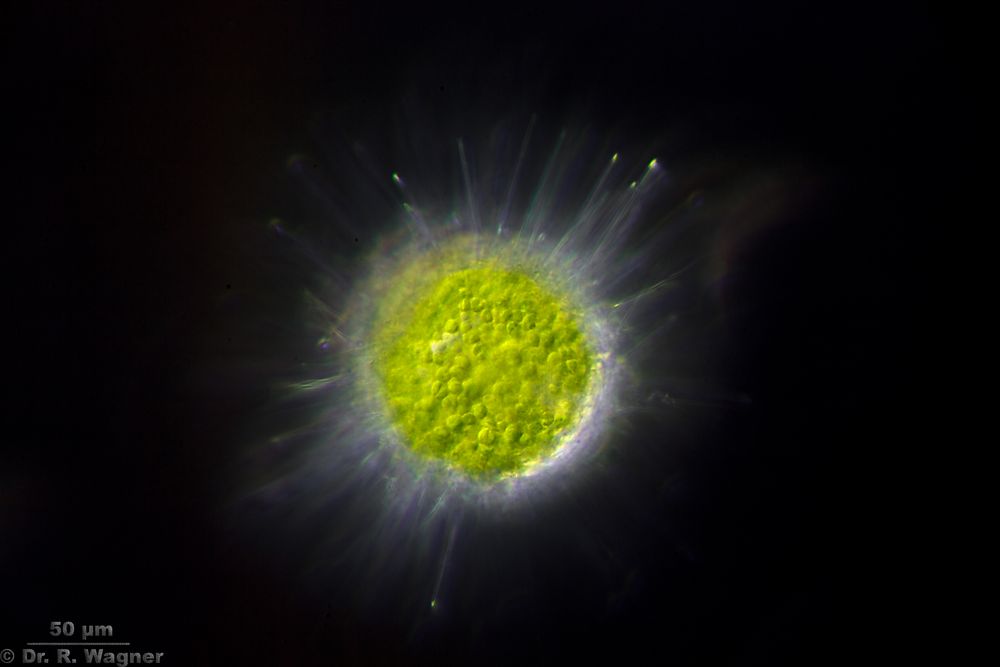
Heterophrys spec. Gardenpond August 2016
Phasecontrast
Heterophrys spec. Gardenpond August 2016
Oblique illum.
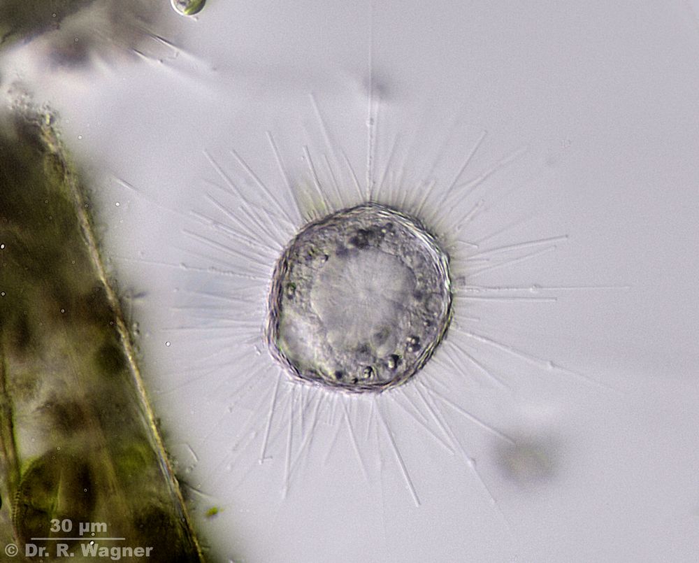
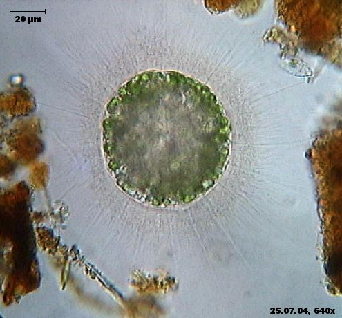
Heterophrys myriapoda, grass-green heliozoan Cologne-Wahn, moor, July 2004
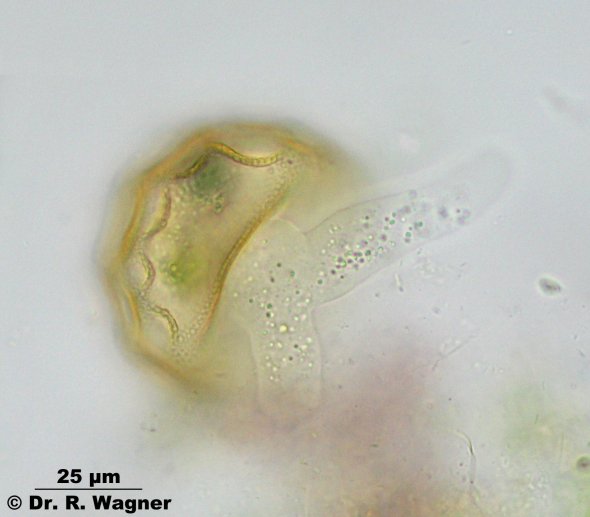
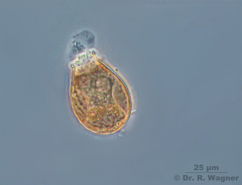
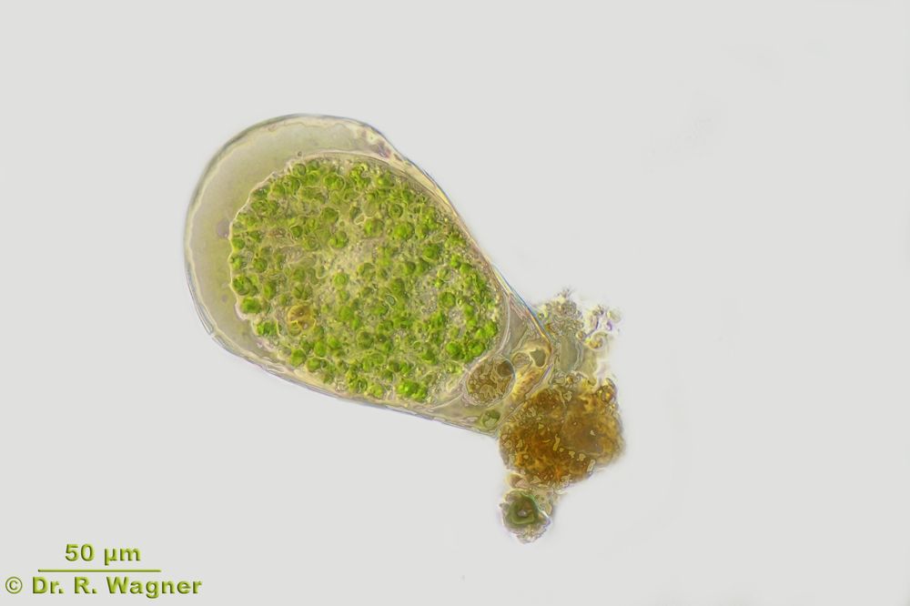
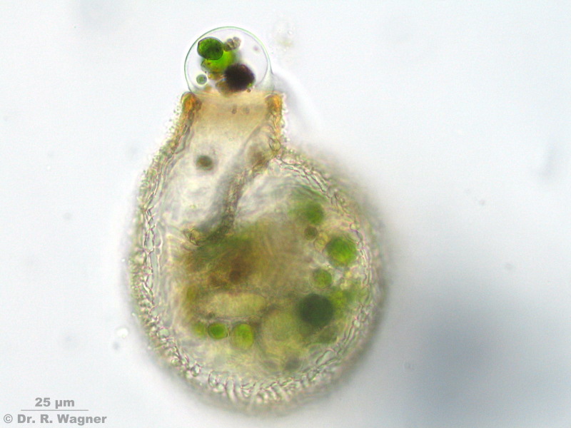
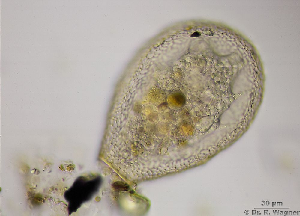
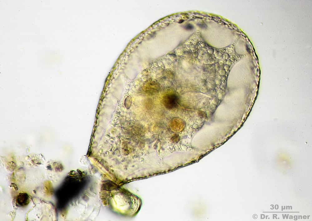
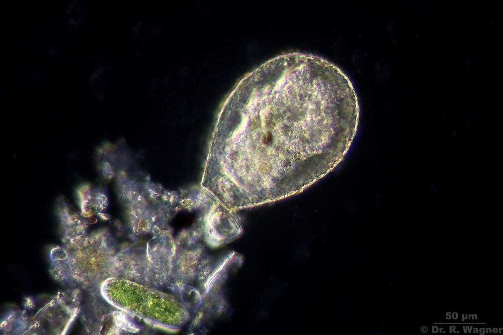
Hyalosphenia papillio Heathland pond near Brüggen, September 2018
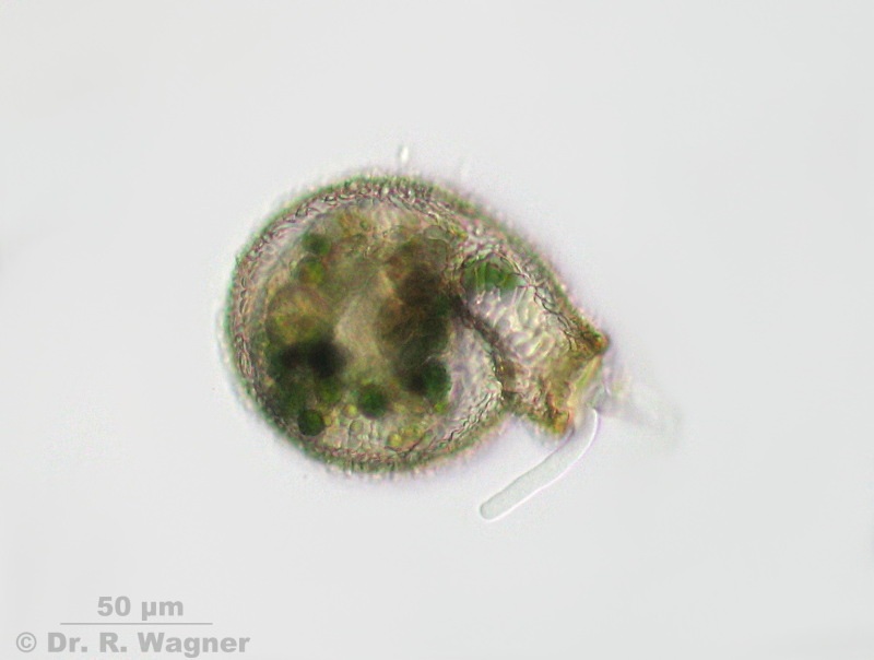
Lesquereusia spiralis Elmpter Schwalmbruch, August 2008
Lesquereusia spiralis builds spiral tests with small silica rods on the surface.
Lesquereusia spiralis Elmpter Schwalmbruch, August 2008
At the test opening there is an excretion-vacuole with indigestible food residuals leaving the shell.
Lesquereusia spiralis Elmpter Schwalmbruch, August 2008
Focus on the silica rods. At the opening there is a lobopodia.
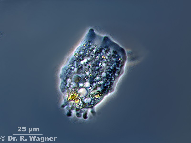
Mayorella spec. Gardenpond
June 2010
Phasecontrast
Mayorella spec. Gardenpond
June 2010
Video on YouTube
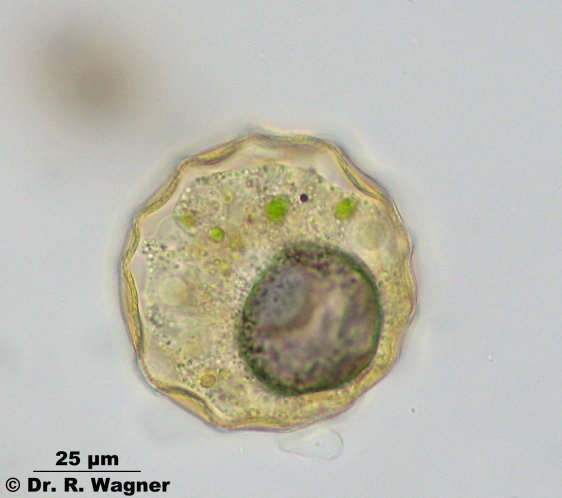
Mayorella viridis with symbiontic green algae
metachaos gratum Hilden heathland, February 2007
Nebela spec. Heathlandpond near Roermond July 2016
Nebela spec. Heathlandpond near Roermond July 2016
Nebela spec. Heathlandpond near Roermond July 2016
Nebela spec. Heathlandpond near Roermond July 2016
Darkfield
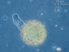
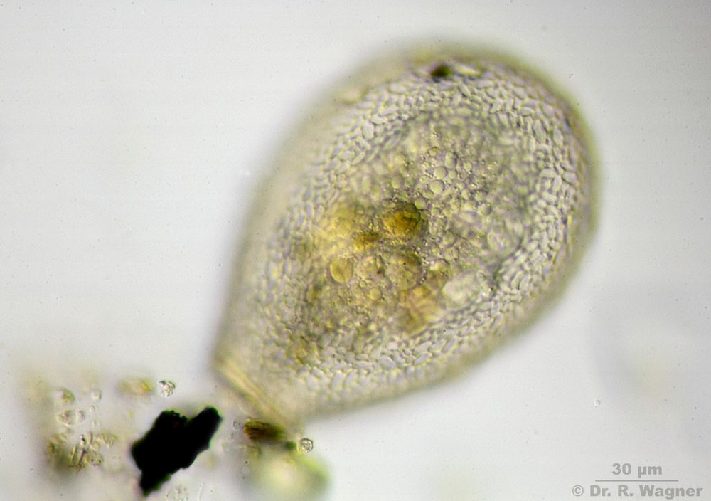
Pseudochlamys arcelloides Ophoven estate, Leverkusen, April 2007
phase contrast
Pseudochlamys arcelloides Ophoven estate, Leverkusen, April 2007
Pseudochlamys arcelloides Ophoven estate, Leverkusen, April 2007
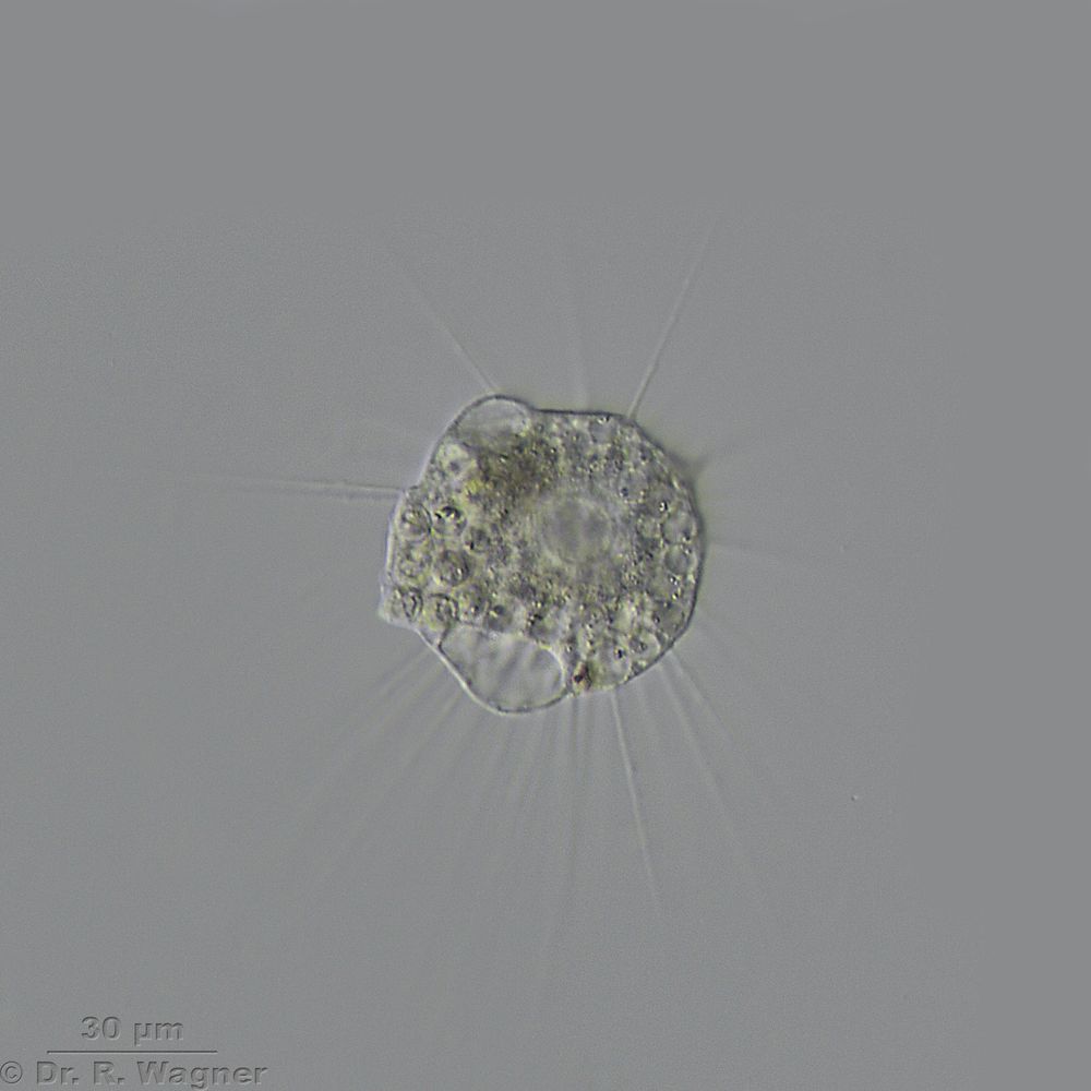
actinophrys sol, the heliozoan Gardenpond September 2016