| preview-picture |
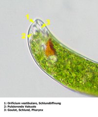
description |
comment |
|
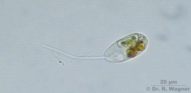
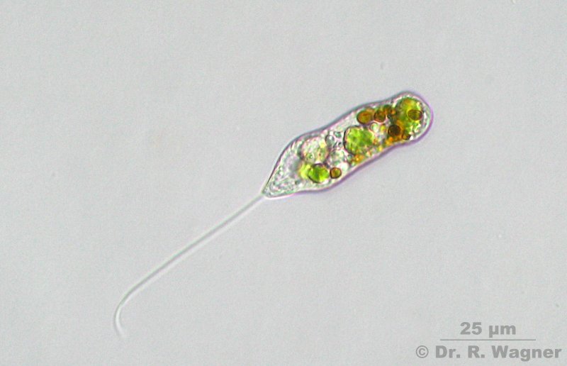
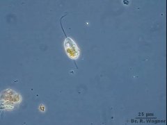
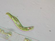
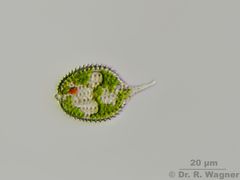
Anisonema spec.
January 2007,
Hilden heathland |
Phasecontrast. The trailing flagellum is of 1,5x cell length, the leading flagellum is of same length as the cell.
The cell-form is not mutable. |
|
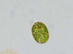
Anisonema spec.
January 2007,
Hilden heathland |
|
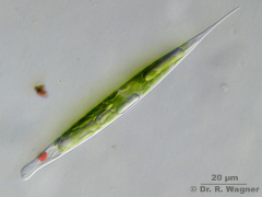 |
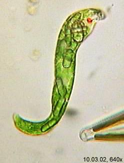
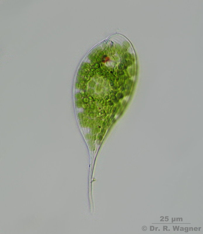
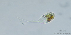
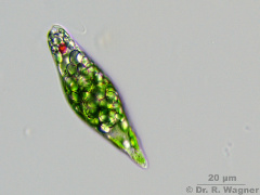
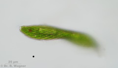
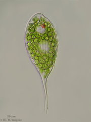
Euglena acus
Gardenpond
February 2010 |
|
|
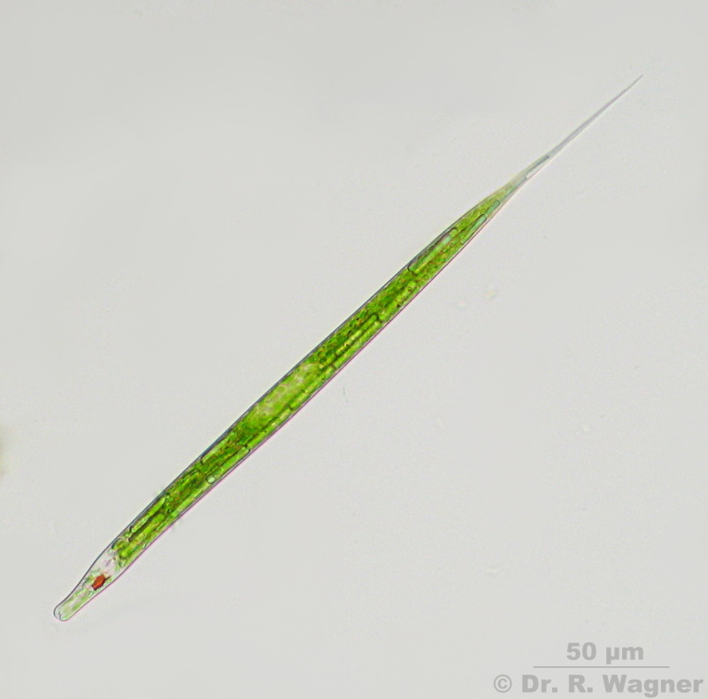
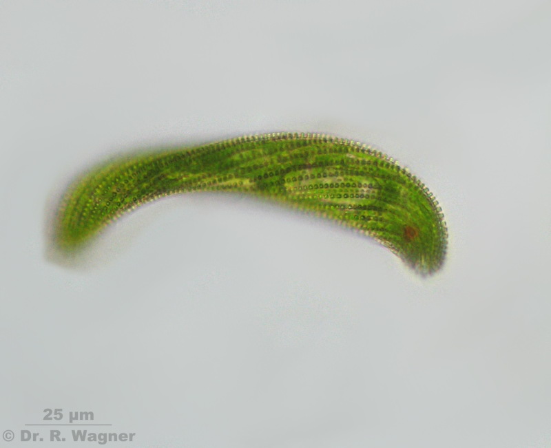
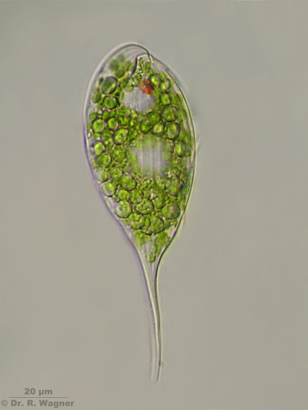
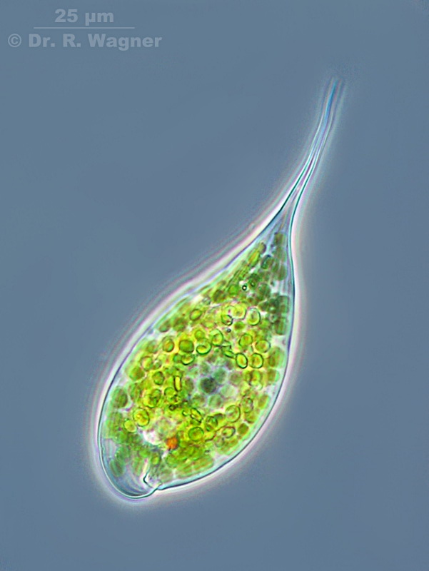
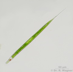
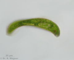
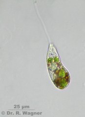
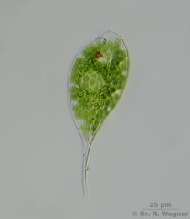
Euglena acus, var. longissima
Heathland pond near Roermond
February 2008 |
|
|
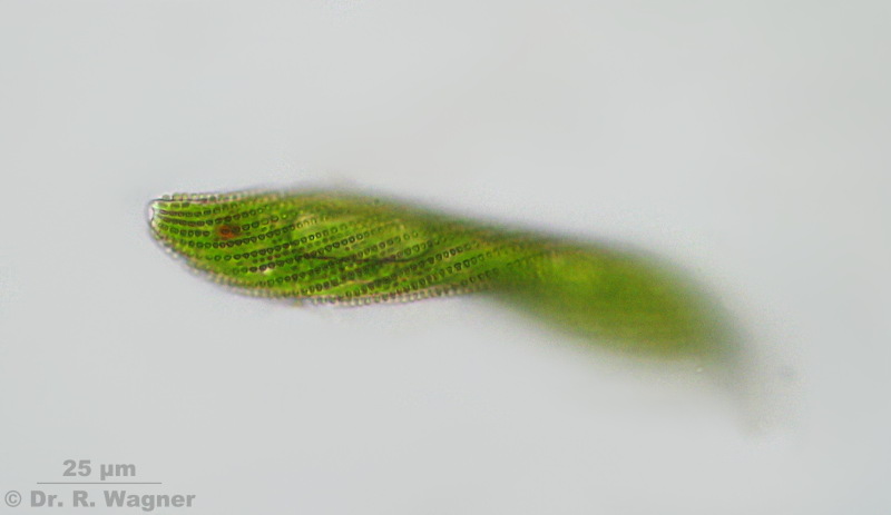
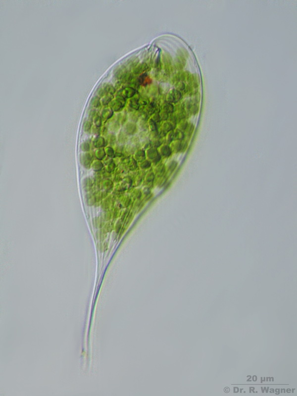
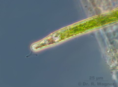
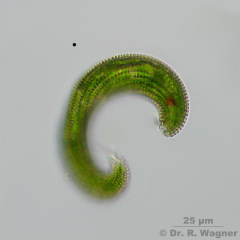
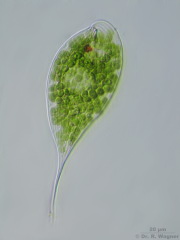
Euglena acus, var. longissima
Heathland pond near Roermond
February 2008 |
Flagellum in phase contrast |

|
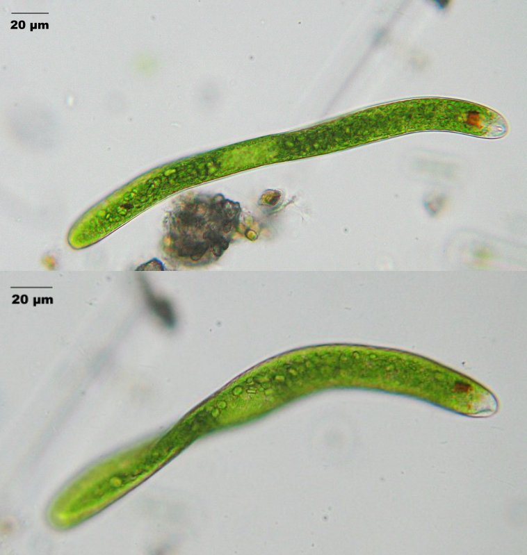
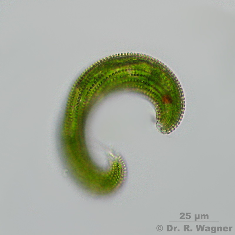
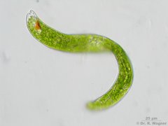
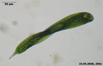
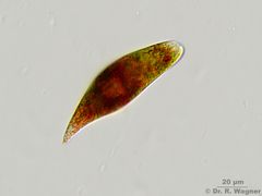
Euglena ehrenbergii
August 2006 |
national horticultural show Duesseldorf |
|
Euglena ehrenbergii
Botanical garden University Düsseldorf, July 2007 |
|
|
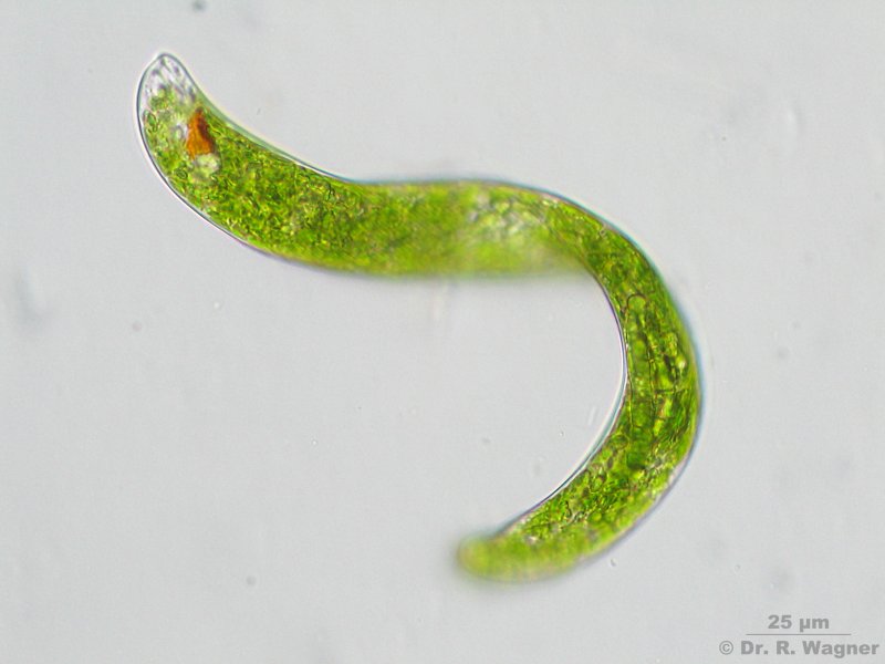
Euglena ehrenbergii
Botanical garden University Düsseldorf, July 2007 |
Detail of the pic above |
 |
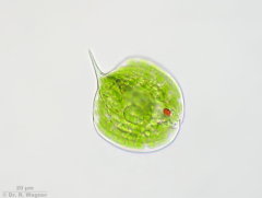
Euglena gracilis
Hilden heathland, March 2009 |
Very active and metabol |
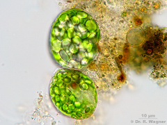 |
Euglena gracilis
Hilden heathland, March 2009 |
Immobile palmella-state |

|
euglena intermedia
March 2002 |
|
|
euglena oxuris
national horticultural show Duesseldorf,
May 2006 |
you can see the red stigma and the distorted shape of this euglena species.
Unfortunately the flagellum could not be resolved.
|
 |
Euglena sanguinea
Hilden heathland
May 2011 |
The red color is caused by the pigment Haematochrom. |
|
Euglena spirogyra
Elfenmeer,
June 2008 |
Typical are the spiral rows of excrescences on the surface-membrane. |
|
Euglena spirogyra
Elfenmeer,
June 2008 |
Focus on the outer membrane, front part with eye spot |
|
Euglena spirogyra
Elfenmeer,
June 2008 |
E. spirogyra is very mutable in form. |
|
euglena spirogyra
Kanzlerberg,
October 2006 |
divx-video, ~3 MB |
 |
Peranema spec.,
Botanical garden University Düsseldorf, July 2007 |
|
 |
Peranema spec.,
Botanical garden University Düsseldorf, July 200 |
|
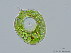 |
Phacus acuminatus
Southpark, Düsseldorf
March 2009 |
Characteristic is the large, ring shaped paramyloncorn in the middle.
|
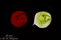 |
Phacus acuminatus
Gardenpond
August 2018 |
Red chlorophyll autofluorescence left, right beneath ordinary illumination |
 |
Phacus acutus
Pietzmoor, Lüneburger Heide
July 2008 |
Periplast with longitudinal stripes; one ring-shaped paramyloncorn; long and straight sting
|
 |
Phacus acutus
Pietzmoor, Lüneburger Heide
July 2008 |
Focus on the striped periplast |
 |
Phacus acutus
Pietzmoor, Lüneburger Heide
July 2008 |
Focus on the striped periplast |
 |
Phacus acutus
Pietzmoor, Lüneburger Heide
July 2008 |
Phasecontrast |
 |
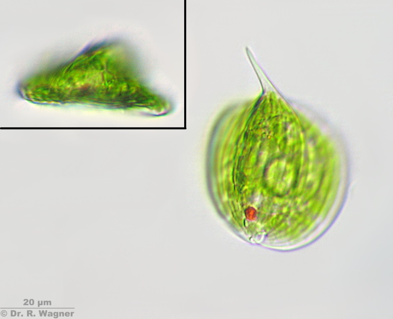
Phacus orbicularis
Southpark, Düsseldorf
August 2008 |
Length: 53 µm, Width: 41 µm. Endsting angular deflected, longitudinal striped periplast,
one central, large paramyloncorn and anothher smaller one, that ist situated excentric to the large one.
|
 |
Phacus orbicularis
Southpark, Düsseldorf
August 2008 |
Focus on the blunt dorsal keel. The inset shows the triangular form of the cell when viewed
in an optical cross-section.
|
 |
Phacus suecicus
pond near in wet meadow near Kempen,
November 2011 |
|
 |
trachelomonas hispida
garden pond,
February 2007 |
|