| preview-picture |
description |
comment |
 |
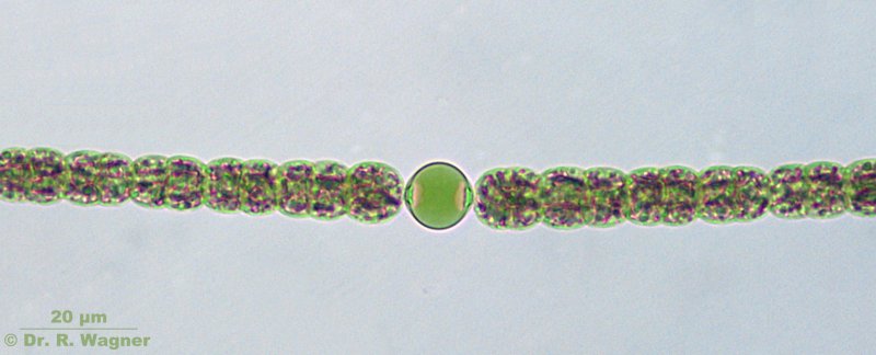
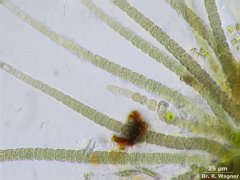
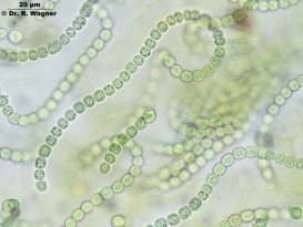
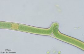
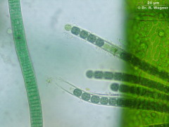
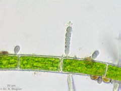
anabaena spec.
botanical garden, university Duesseldorf
August 2006 |
The picture shows an anabaena spec. with cells in different cell-division stages.
You can see also the somewhat bigger heterocystis in the middle of the filament.
Here the nitrogen processing takes place. At the contact points with the "normal" cells there
are two light scattering granules visible. These are the so called pole-bodies.
Through these the nitrogen-assimilates are transported to all other cells.. |
 |
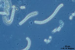
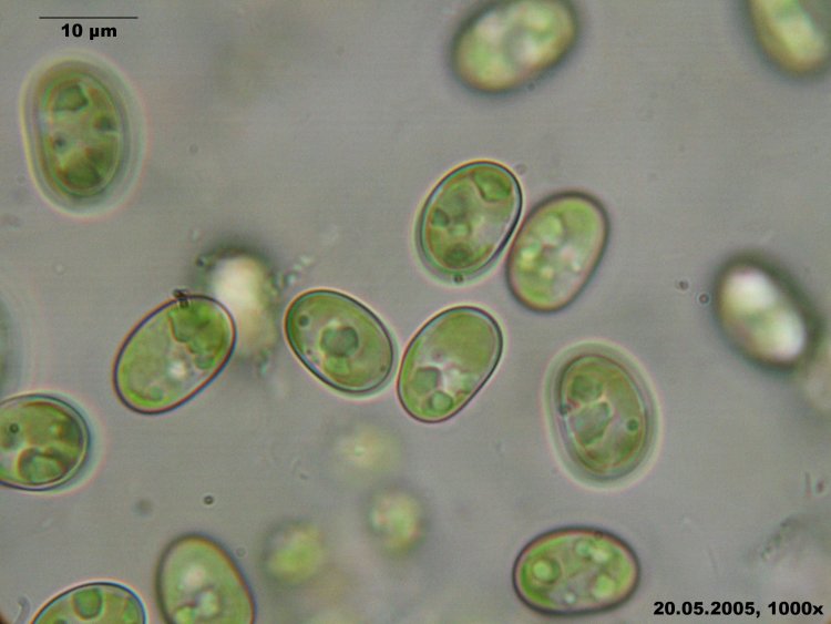
aphanothece spec.
garden pond
May 2005 |
|
 |
aphanothece spec.
Rhine bank near Urdenbach
Oktober 2005 |
|
 |
Chamaesiphon spec.
Aquariumf
February 2008 |
|
 |
Chamaesiphon spec.
Aquariumf
February 2008 |
|
 |
Chroococcus multicoloratus
Mucilaginous film from an old railwaystation wall
January 2016 |
|
 |
Chroococcus turgidus
gardenpond
August 2016 |
|
 |
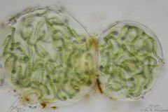
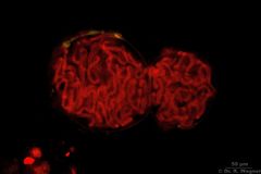
Coelosphaerium spec.
Gardenpond
August 2016 |
|
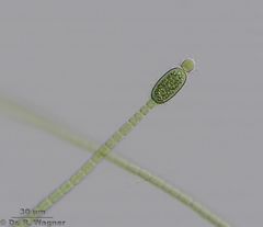 |
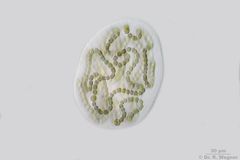
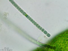
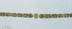
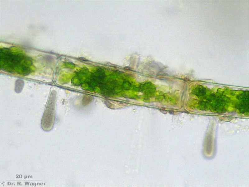
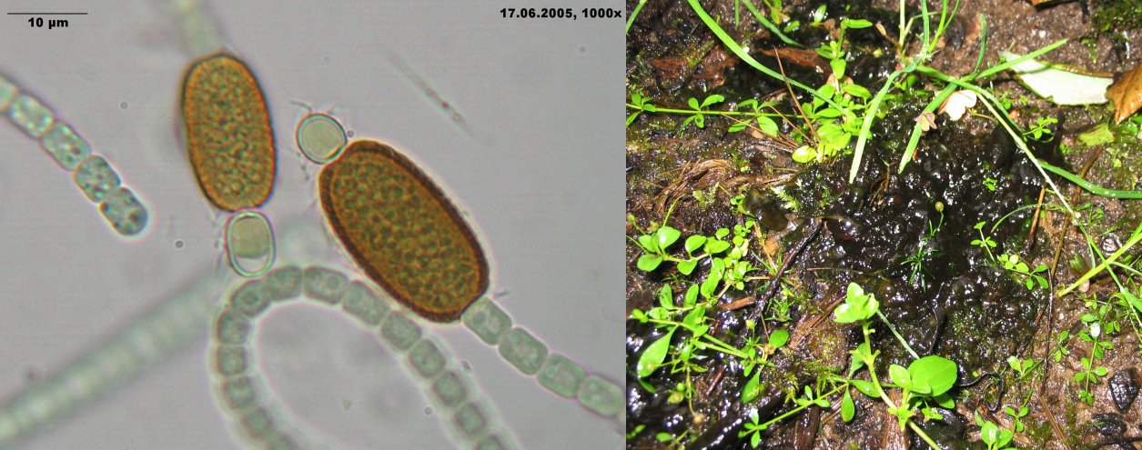
Cylindrospermum
garden pond, bank
July 2016 |
At the end of the filament an longish akinet and a round heterocyst |
|
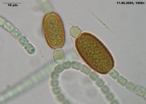
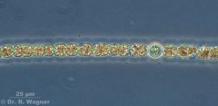
cylindrospermum spec.
garden pond, bank
Juni 2005 |
At the end of the filament an longish akinet and a round heterocysta.
The right picture shows the habitus as seen with your eyes. |

|
gleothece linearis
Cologne-Wahn, moor
December 2006 |
Single cells linear arranged in a common gelatin |
 |
Gloeocapsa spec.
Slime on a wall at a railwaystation
January 2016 |
Single cells in common gelatin |
 |
Gloeocapsa spec.
Slime on a wall at a railwaystation
January 2016 |
Single cells in common gelatin |
 |
Gloeocapsa spec.
Slime on a wall at a railwaystation
January 2016 |
Single cells in common gelatin |
|
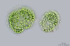
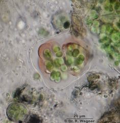
Gloeotrichia echinulata
Seethaler lake near Tamsweg, Austria
September 2007 |
Overall view |
|
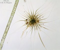
Gloeotrichia echinulata
Seethaler lake near Tamsweg, Austria, Österreich
September 2007 |
Trichomes end in in long, colorless hairs |
|
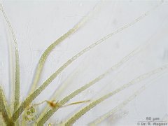
Gloeotrichia echinulata
Seethaler lake near Tamsweg, Austria, Österreich
September 2007 |
|
|
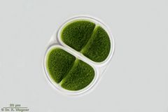
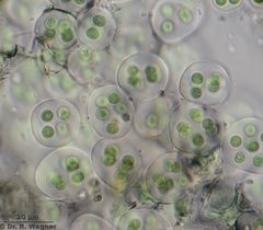
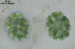
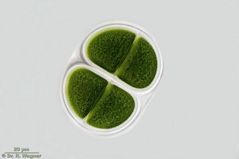
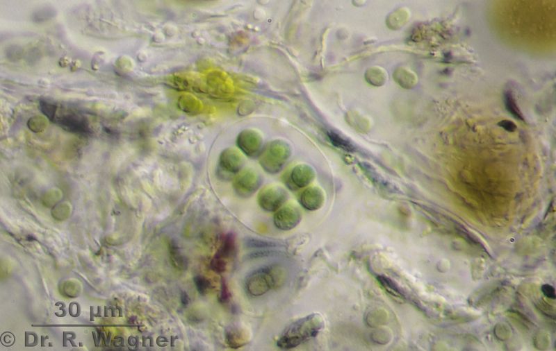
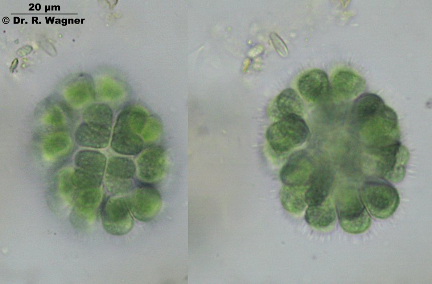
gomphospaeria aponina
botanical garden, university Duesseldorf
Dezember 2006 |
|
|
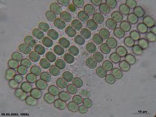
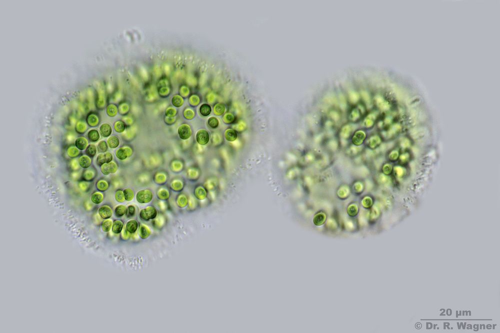
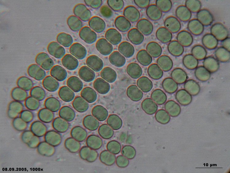
merismopedia elegans
north sea coast, Emden
September 2005 |
|
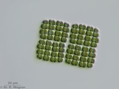 |
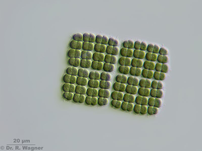
Merismopedia spec
Small pond near Hilden
June 2012 |
|
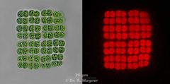 |
Merismopedia spec.
Birgelen, virgin forest
April 2013 |
Brightfield and Chlorophyll autofluorescence in 365 nm UV-excitation
|
-azollae_K.jpg)
|
nostoc-(anabaena)-azollae
river Erft near Frimmersdorf
April 2003 |
The right picture shows the algae nostoc-(anabaena)-azollae. The pictures to the left show
the fern azolla filiculoides. It forms a symbiosis with the cyanobactrium which is living
in caves formed by the ferns leaves. |
-azollae-HF_K.jpg)
|
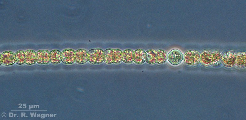
Nostoc-(Anabaena)-azollae
garden pondh
April 2007 |
The symbiontic cyanobacteriume of the fern Azolla filiculoides.
Filament with heterocysts, at the contact-dots of the heterocyst we find the light scattering
pole-bodies. |
-azollae-PH_K.jpg)
|
Nostoc-(Anabaena)-azollae
garden pond
April 2007 |
The symbiontic cyanobacteriume of the fern Azolla filiculoides in phase contrast. |
 |
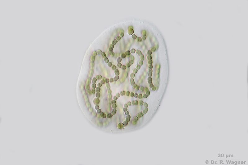
Nostoc linckia
gardenpond
July 2015 |
|
 |
Nostoc linckia
Heathland pond near Roermond
July 2016 |
|
 |
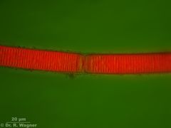
Nostoc linckia
Heathland pond near Roermond
July 2016 |
Chlorophyll Autofluorescence |

|
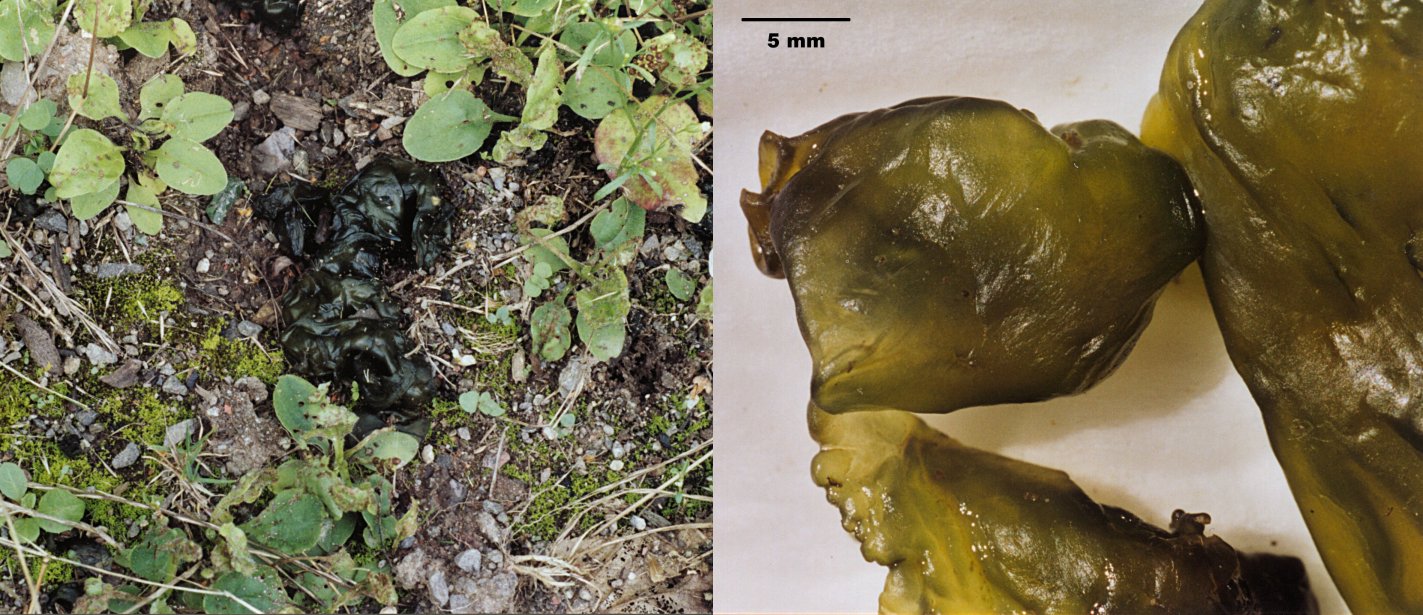
nostoc commune
wayside of a little park in Benrath
August 2004 |
macrofoto, habitus |
|
nostoc commune
savaged site in Duesseldorf Urdenbach
Dezember 2006 |
|
|
nostoc commune
savaged site in Duesseldorf Urdenbach
Dezember 2006 |
same as above, but with phase-contrast |
 |
Oscillatoria spec.
Small pond near Hilden
June 2012 |
|
 |
Oscillatoria spec.
Small pond near Hilden
June 2012 |
Chlorophyll Autofluorescence |
|
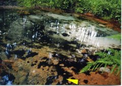
The Schwinde-spring, Lüneburger Heide
Juliy 2008 |
This picture shows the habitat of the folowing 3 pictures. The yellow arrow marks
the large, black, macroscopically visible beds of Oscillatoria limosa.
|
|
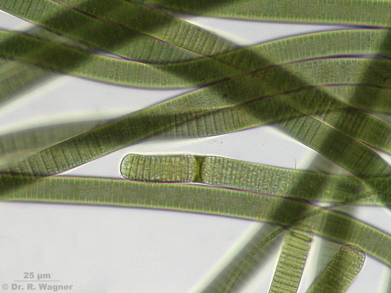
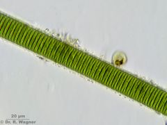
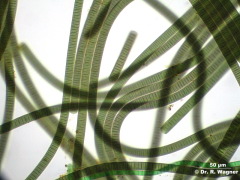
Oscillatoria limosa
Schwinde-spring, Lüneburger Heide
July 2008 |
Filaments are 15 µm width, cellength is 4 µm, filaments are non-bransched and have a small gelantineous shell
|

|
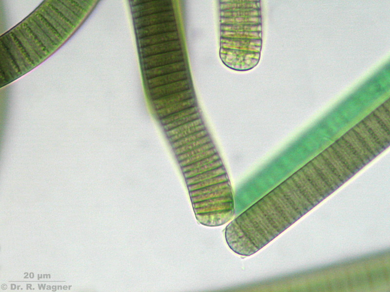
Oscillatoria limosa
Schwinde-spring, Lüneburger Heide
July 2008 |
Formation of hormogonias
|

|
Oscillatoria limosa
Schwinde-spring, Lüneburger Heide
July 2008 |
The ending-cells are flatly rounded
|

|
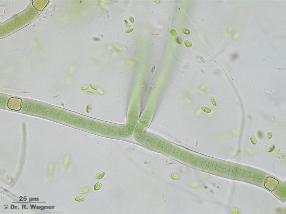
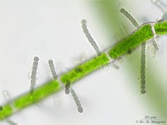
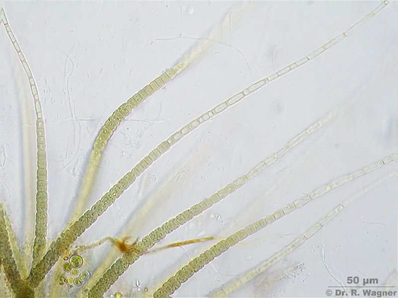
scytonema spec.
Cologne-Wahn, moor
January 2007 |
The genus Scytonema shows false branchings. This means, that the branch is generated from two
independent filaments and not by the cells of a single filament. Furthermore there are three
heterocysts visible. Heterocysts are special cells that are able to synthesize organic (nitrogen)
compounds from the nitrogen in the air. Also visible is the common mucous borderline. |
|
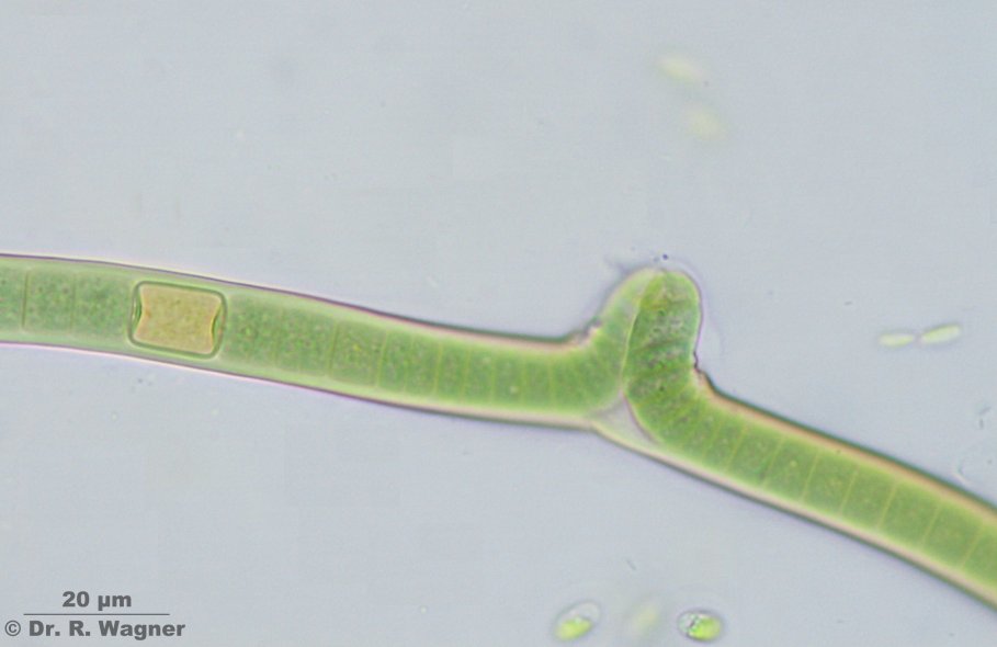
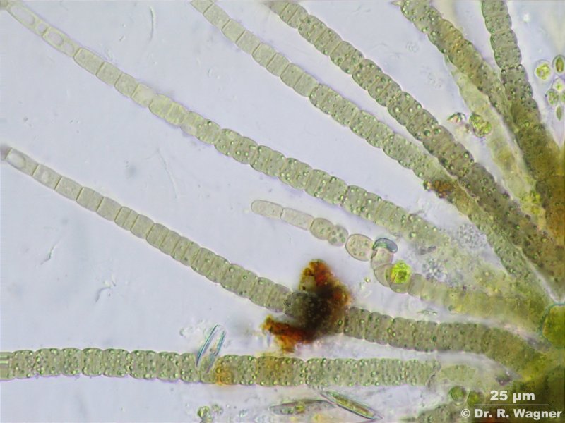
scytonema spec.
Cologne-Wahn, moor
January 2007 |
early state of branching |
|
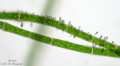
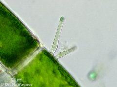
Stichosiphon sansibaricus
Aquarium
February 2008 |
Epiphytic species |
|
Stichosiphon sansibaricus
Aquarium
February 2008 |
Epiphytic species |
|
Stichosiphon sansibaricus
Aquarium
February 2008 |
Epiphytic species |
|
Stichosiphon sansibaricus
Aquarium
February 2008 |
Epiphytic species |
|
Stichosiphon sansibaricus
Zoo Krefeld, Tropical center
August 2008 |
Epiphytic on Cladophora |
|
Stichosiphon sansibaricus
Zoo Krefeld, Tropical center
August 2008 |
Epiphytic on Cladophora |
|
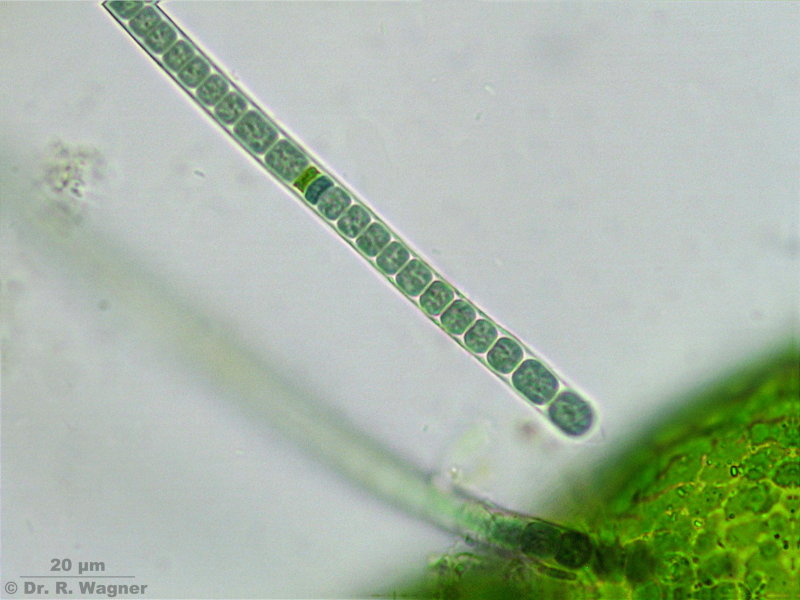
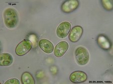
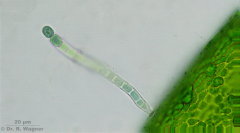
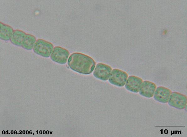
Trichormus catenulus var. affinis
Pond near Hilden
June 2007 |
|
|
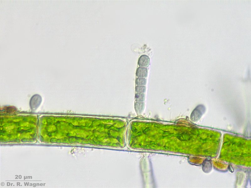
Trichormus catenulus var. affinis
Pond near Hilden
June 2007 |
In phase-contrast the small gelantineous envelope becomes visisble |


-azollae.jpg)
-azollae-HF.jpg)
-azollae-PH.jpg)