| preview-picture |
description |
comment |

|
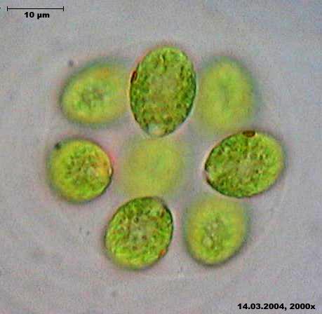
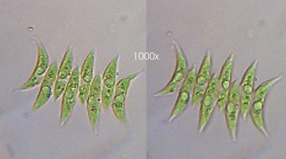
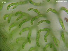
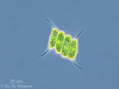
ankistrodesmus spiralis
renaturated clay pit near Bergisch-Gladbach
June 2005 |
|
 |
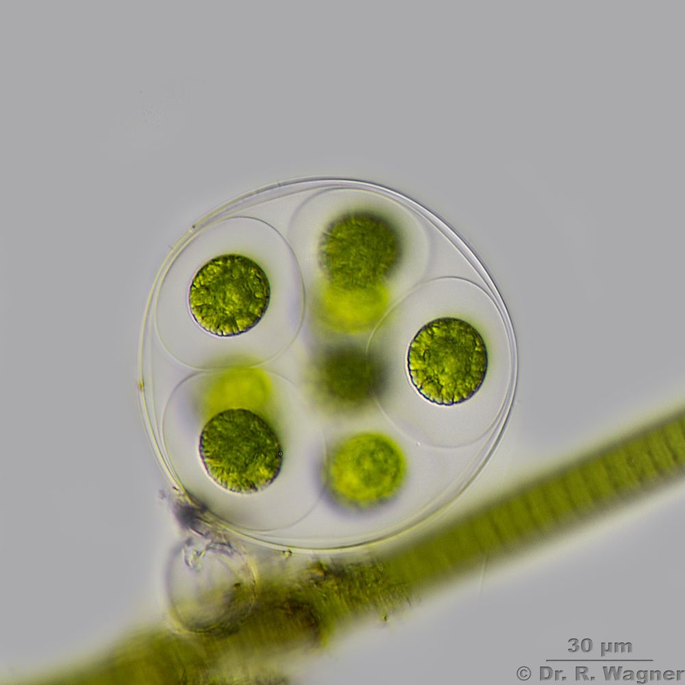
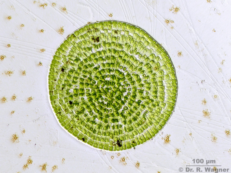
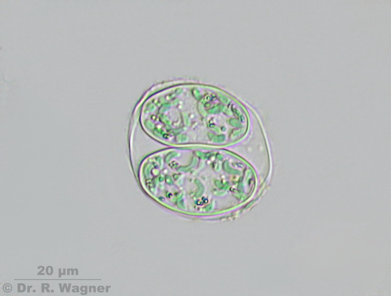
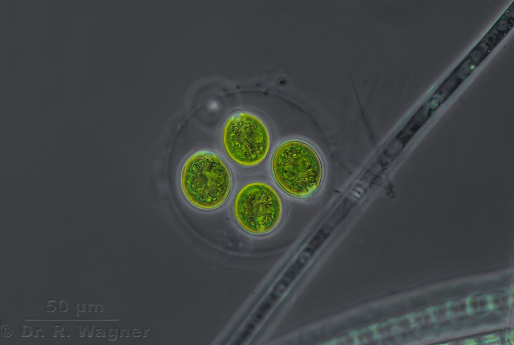
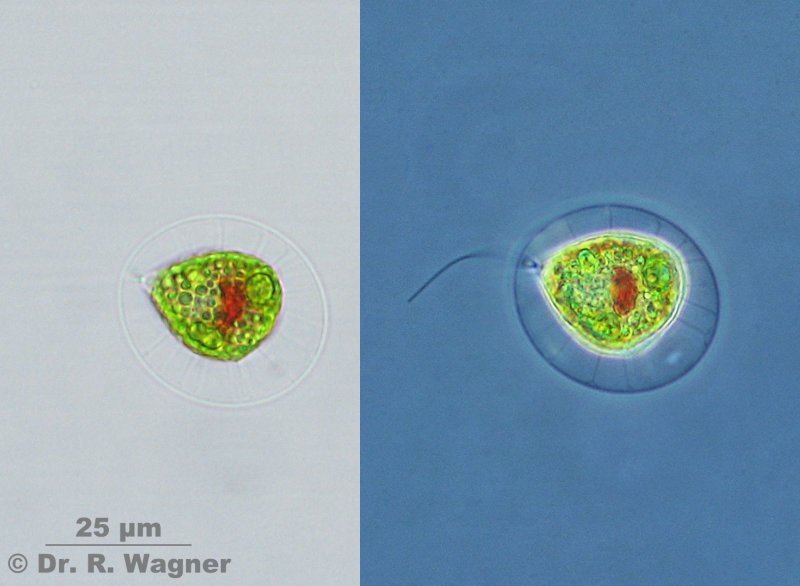
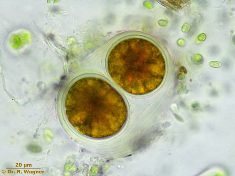
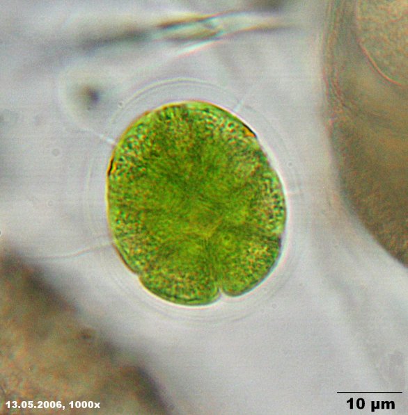
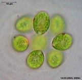
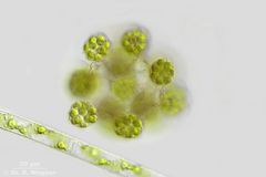
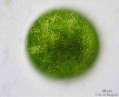
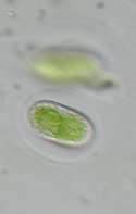
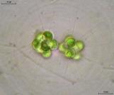
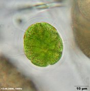
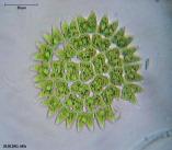
Asterococcus superbus
Gardenpond
September 2016 |
You can see the concentric layered shell-membranes |
 |
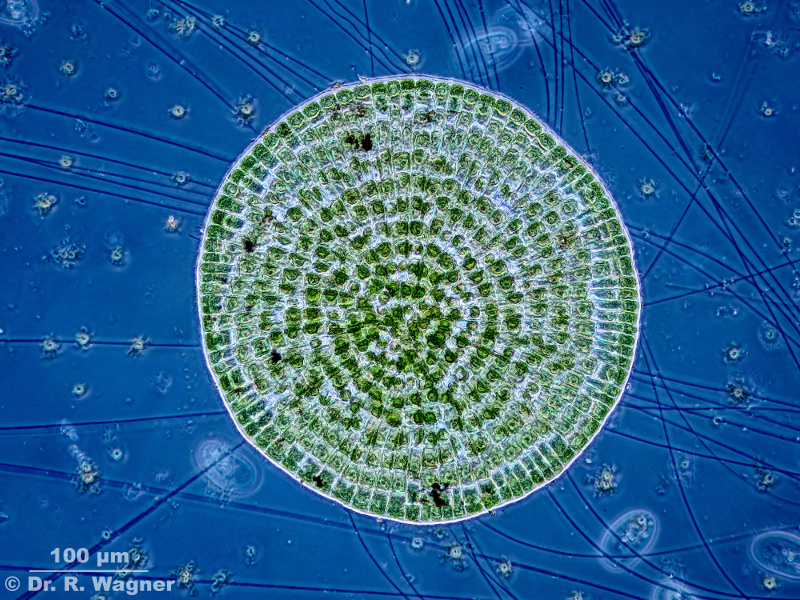
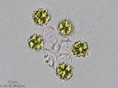
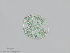
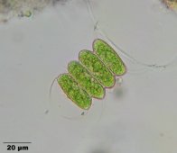
Asterococcus superbus
Gardenpond
September 2016 |
Phasecontrast |
|
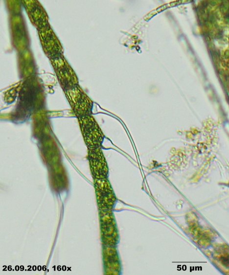
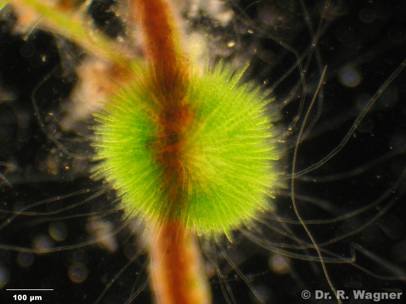
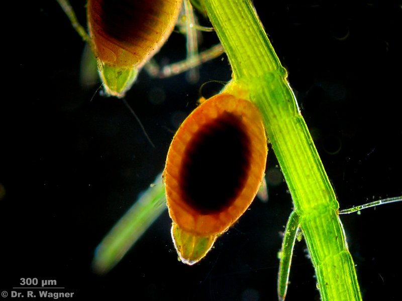
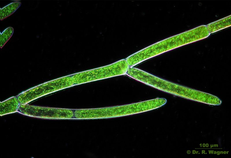
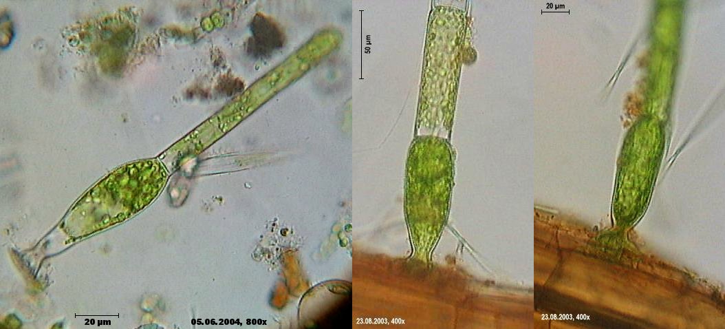
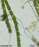
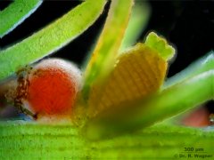
Bulbochaete spec.
national horticultural show Duesseldorf
September 2006 |
typical are the long hairs that have an onion-like form at the basis |

|
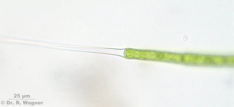
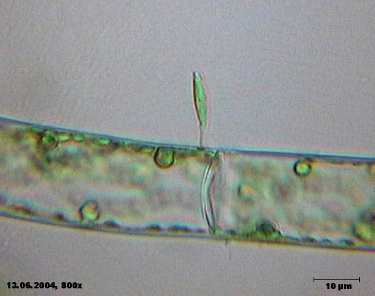
Bulbochaete spec.
botanical garden, university Duesseldorf
May 2007 |
Hair at the end of filament |
|
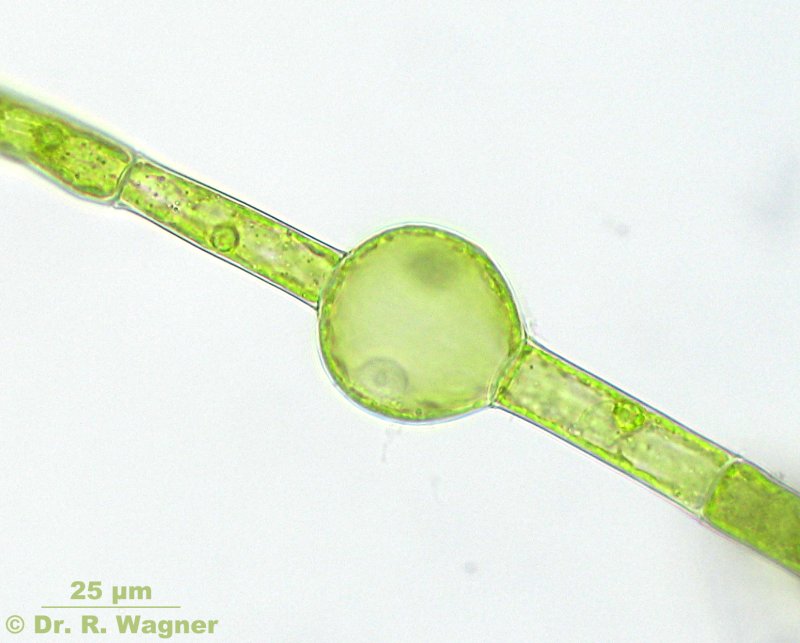
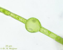
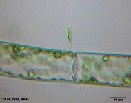
Bulbochaete spec.
botanical garden, university Duesseldorf
May 2007 |
Oogonium with ovum |
|
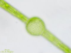
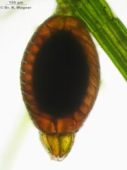
Bulbochaete spec.
botanical garden, university Duesseldorf
May 2007 |
Cell-wall of the oogonioum |
 |
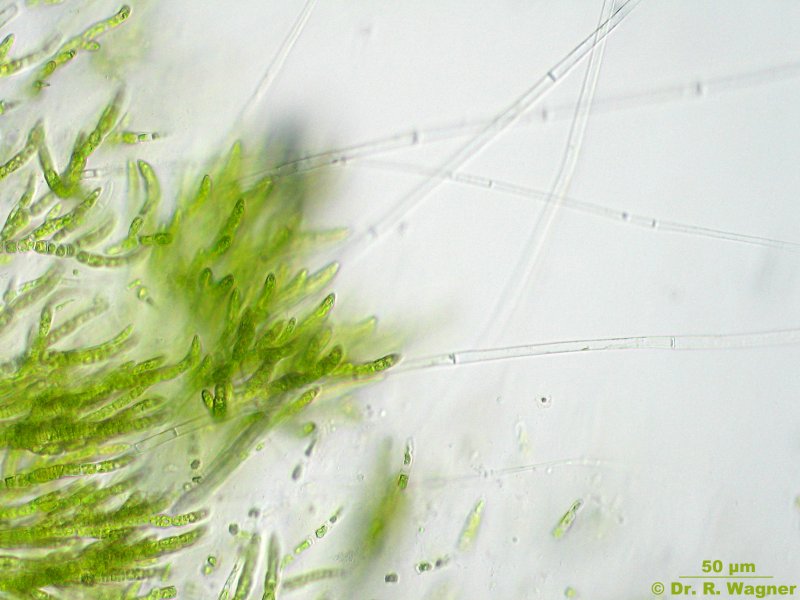
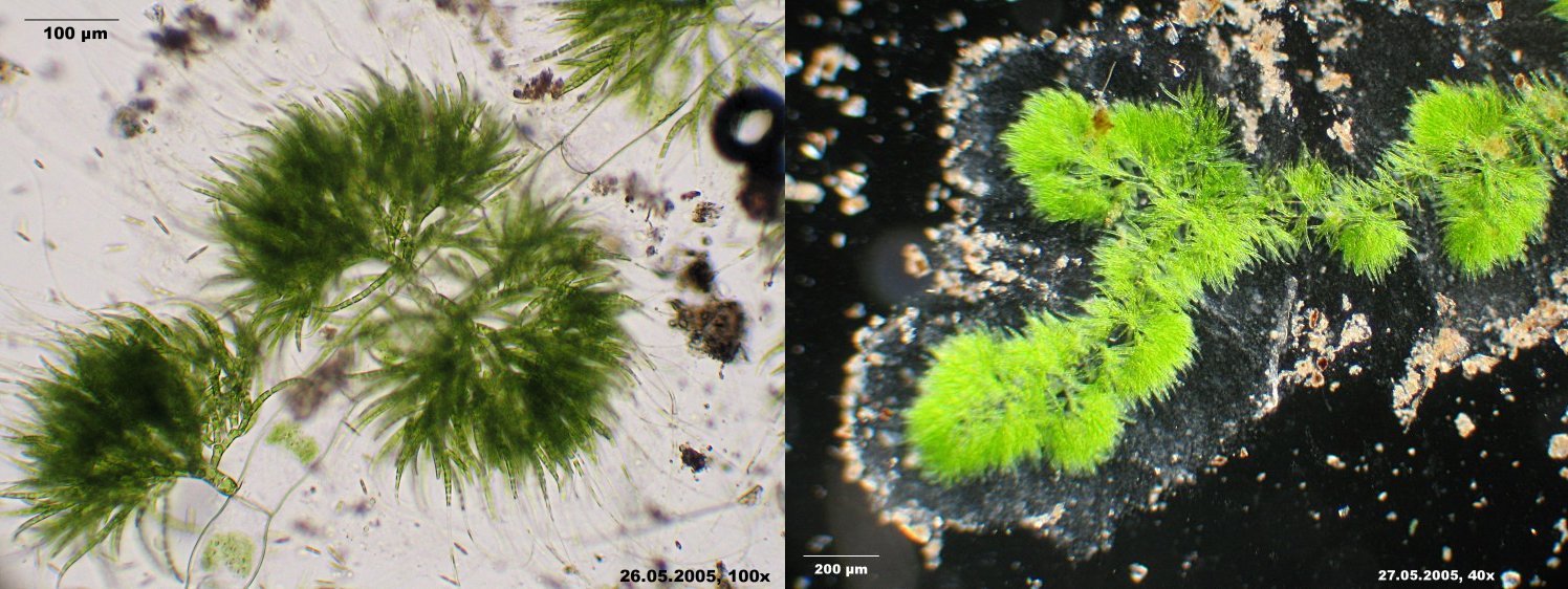
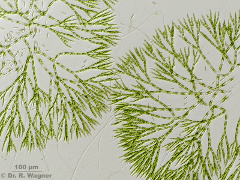
Chaetophora spec.
garden pond
April 2005 |
Habitus, some filaments end in a long "hair" without chloroplasts.
Stereomicroscope |
 |
chaetophora elegans
garden pond
April 2007 |
each hair consists of single, empty cells |
 |
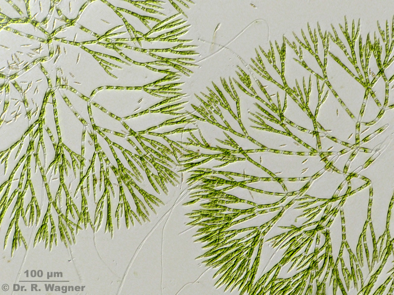
Chaetophora spec.
garden pond
May 2010 |
Chaetophora forms branched filaments |
|
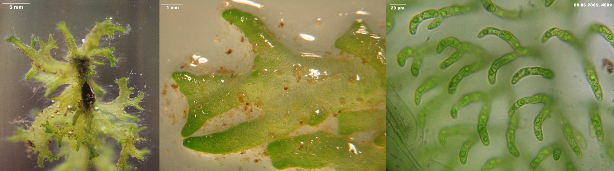
chaetophora incrassata
botanical garden, university Duesseldorf
June 2005 |
similar to Ch. elegans, however macroscopically it forms a branches, lobated gelatin. |
|
Chara spec.
Eskesberg in Wuppertal
August 2007 |
This is a monoecious species. Oogonium (right) and antheridium (left, orange colored) appear on one individual. |
|
Chara spec.
Garden pond
August 2007 |
This is a dioecious species. Oogonium and antheridium appear on two different plants;
here a female with an oogonium individual is shown. |

|
Chara spec.
Garden pond
August 2007 |
This is a dioecious species. Oogonium and antheridium appear on two different plants;
here a female with an oogonium individual is shown. |
|
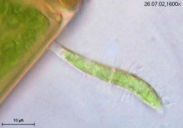
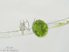
characium, species
old Emscher, Emscherpark
June 2004 |
|

|
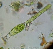
characium, species
Unterbacher pond
July 2002 |
|

|
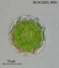
zygote of chlamydomonas species
garden pond
April 2003 |
|
|
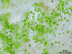
chlorella vulgaris
garden pond
December 2006 |
shown here as the symbiontic form that occurs in stentor polymorphus. The two circles mark the
typical forms of the chloroplast. |
|
chlorococcum spec.
garden pond
March 2004 |
Division in daughter-cells |

|
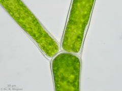
Cladophora glomerata
Waterfall Neandertal
March 2008 |
Cladophora consists of branched filamentsMicroscopy, Stereomicroscope, Microfototo |
 |
Cladophora glomerata
Waterfall Neandertal
March 2008 |
Branching |

|
Cladophora glomerata
Waterfall Neandertal
March 2008 |
A new branching evolves through a protuberance under a transverse cell wand |
 |
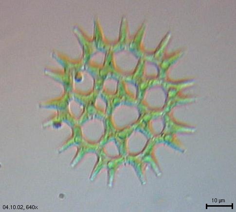
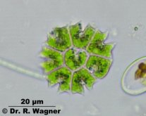
Coelastrum astroideum
Small pond near Hilden
May 2011 |
|
 |
coelastrum microporum
Eselsbach near the Unterbacher pond
August 2003 |
|
 |
Coelastrum reticulatum
Gardenpond, Lower Rhine region
July 2016 |
Coelastrum reticulatum has been brought to Europe from the tropics
at the beginning of the 20th. century. |
 |
Coelastrum reticulatum
Northpark, Düsseldorf
June 2011 |
Coelastrum reticulatum has been brought to Europe from the tropics
at the beginning of the 20th. century. |
|
coelastrum sphaericum
botanical garden university Duesseldorf
August 2006 |
4 pictures stacked with CombineZ5 |
 |
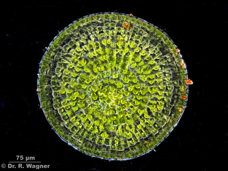
Coleochaete orbicularis
Garden pond
July 2010 |
24 pictures stacked with Zerene stacker |
 |
Coleochaete orbicularis
Garden pond
July 2010 |
Phase contrast, 10 pictures stacked with Zerene stacker |
 |
Coleochaete orbicularis
Garden pond
July 2010 |
Dark field, 20 pictures stacked with Zerene stacker |
|
dictyosphaerium ehrenbergianum
pond near the Wupper dam
May 2006 |
|

|
draparnaldia glomerata
garden pond
May 2005 |
In the dark-field-picture to the left you can see the highly viscous
gelantin that surrounds draparnaldia. |
|
Eremosphaera viridis
Seethaler lake near Tamsweg, Austria
September 2007 |
green moor-ball, large form |

|
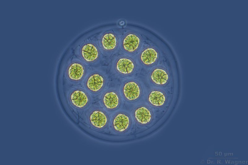
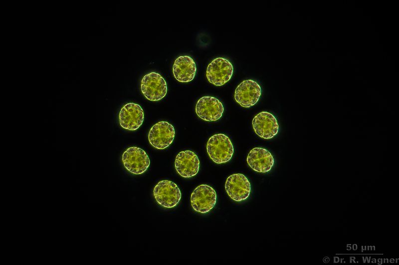
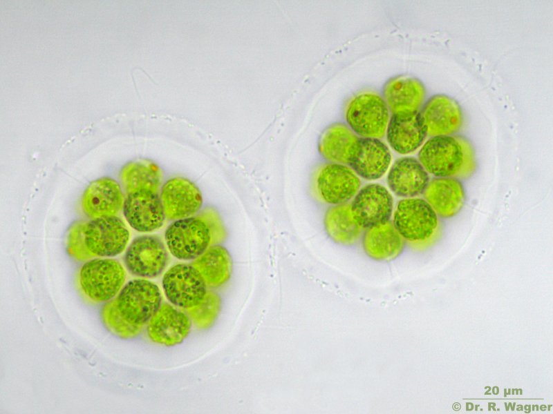
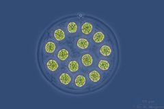
eudorina elegans
Köln-Wahn moor, pond,
May 2002 |
|

|
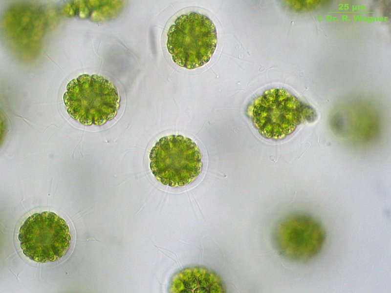
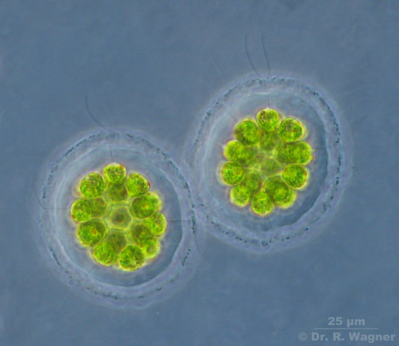
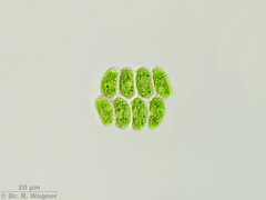
eudorina spec
botanical garden university Duesseldorf,
August 2006 |
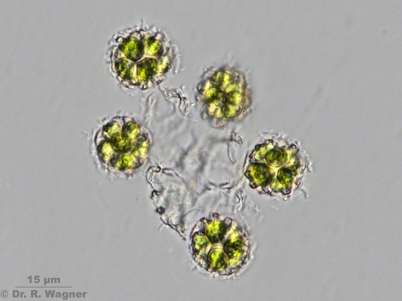
here we see the relatively unusual 8-celled form of eudorina |
|
Glaucocystis spec.
Heathland pond near Roermond,
February 2008 |
Contains symbiontic cyanobacterias instead of a green chloroplast |
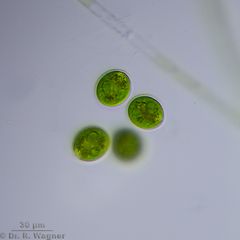 |
Gloeococcus spec.
Heathlandpond near Roermond,
June 2015 |
|
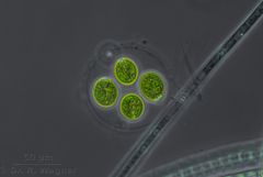 |
Gloeococcus spec.
Heathlandpond near Roermond,
June 2015 |
Phasecontrast |
|
gloeocystis ampla
border of garden pond,
September 2006 |
left: habitus, outside of the pond, between moss
middle: colony of several cells,
right; single cell in layered gelatin |

|
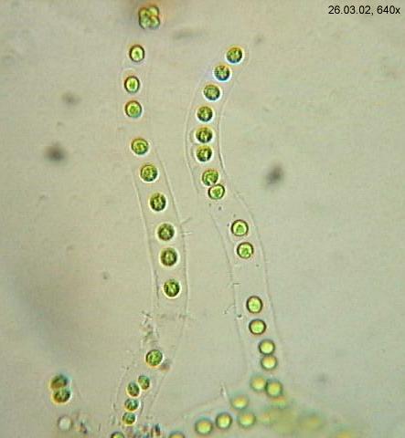
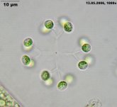
gonium pectorale
garden pond,
April 2002 |
right picture: early state of reproduction |

|
gonium pectorale
garden pond,
April 2003 |
late state of reproduction: it is visible, that from each single cell a complete
16-celled coenobia is generated. |
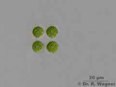 |
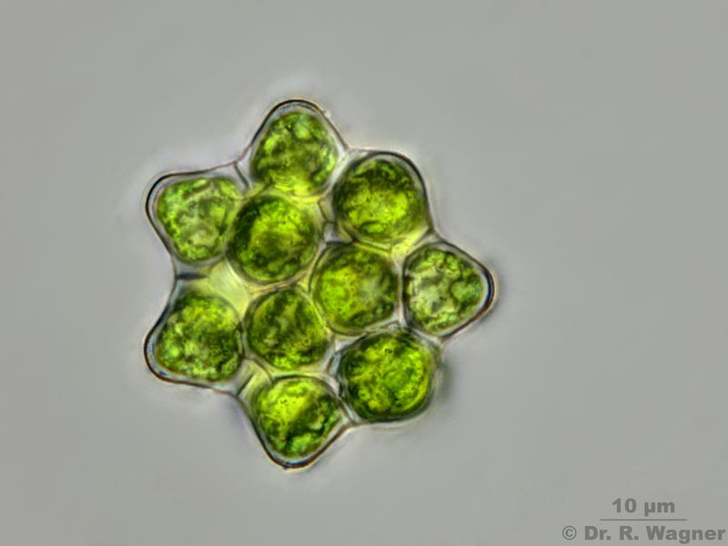
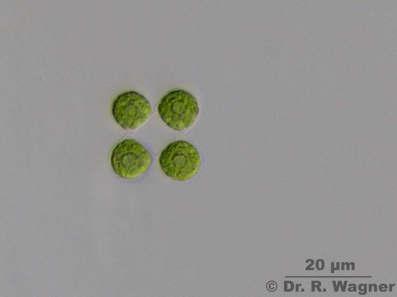
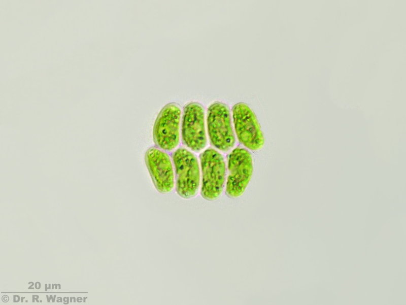
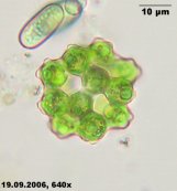
Gonium sociale
Uni Düsseldorf, Botanical garden,
February 2011 |
Always 4-celled, cells quadratically ordered and connected via a small papillae, central pyrenoid, eye-spot.
|

|
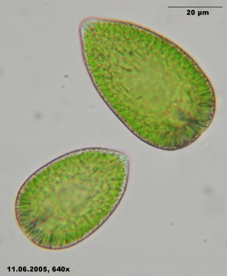
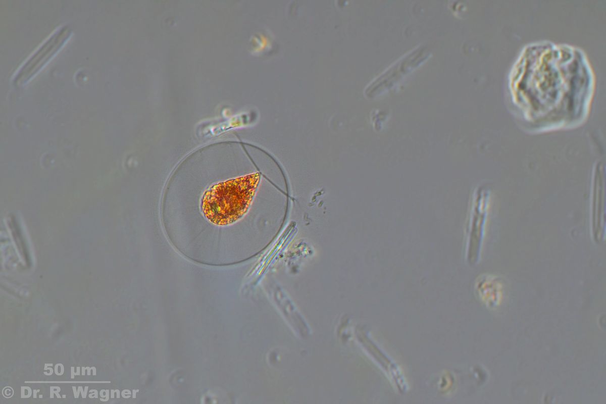
gonyostomum semen
Elfenmeer, a heathland pond
June 2005 |
|
 |
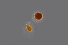
Haematococcus pluvialis
Birds bath, Düsseldorf
September 2021 |
Phasecontrast |
 |
Haematococcus pluvialis
Birds bath, Düsseldorf
September 2021 |
Phasecontrast |

|
haematococcus pluvialis
eaves gutter, Duesseldorf
January 2007 |
|

|
haematococcus spec.
eaves gutter, Duesseldorf
January 2007 |
aplanospores |

|
haematococcus spec.
Köln Wahn moor, December 2006 |
state of cell division |
|
microspora spec.
Köln Wahn moor, pond
April 2006 |
|

|
microspora spec.
atmophytic in a gelantin between mosses, garden
January 2007 |
|

|
microspora spec.
atmophytic in a gelantin between mosses, garden
January 2007 |
|
|
micractinium pusilium
little creek near Roermond
October 2002 |
|
|
nephrocytium lunatum
garden pond
July 2002 |
|
 |
Oedogonium spec.
Garden pond
August 2008 |
Left to the oogonium there are 2 males . One is already empty, the other one cotains still the spermatozoides.
|
 |
Oedogonium spec.
Garden pond
August 2008 |
The male is tightened via a rhizoid at the cellwall.
|
 |
Oedogonium spec.
Garden pond
August 2008 |
Cap cell. These caps are formed during the cell division.
|
|
oedogonium spec.
garden pond
June 2004 |
young filament with rhizoid |

|
palmodictyon viride
botanical garden university Duesseldorf,
March 2002 |
|
|
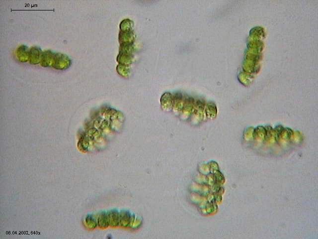
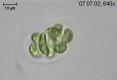
pandorina morum
pond near the Wupper dam,
May 2006 |
|
 |
Pandorina morum
Gardenpond,
July 2015 |
Phasecontrast, colony in asexual reproduction. Each of the usuallay 16 single colony-members
is generating a complete 16-celled daughter-colony. |
 |
Pandorina morum
Gardenpond,
July 2015 |
Darkfield, colony in asexual reproduction. Each of the usuallay 16 single colony-members
is generating a complete 16-celled daughter-colony. |
 |
Pandorina morum
Gardenpond,
July 2015 |
Colony in asexual reproduction. Each of the usuallay 16 single colony-members
is generating a complete 16-celled daughter-colony. |

|
Pandorina morum
Southpark Duesseldorf,
April 2007 |
Each colony is surrounded by a gelantine shell. |

|
Pandorina morum
Southpark Duesseldorf,
May 2007 |
Each cell with 2 flagella |

|
Pandorina morum
Southpark Duesseldorf,
May 2007 |
Cells are wedge-shaped with the flat side exterior. |
|
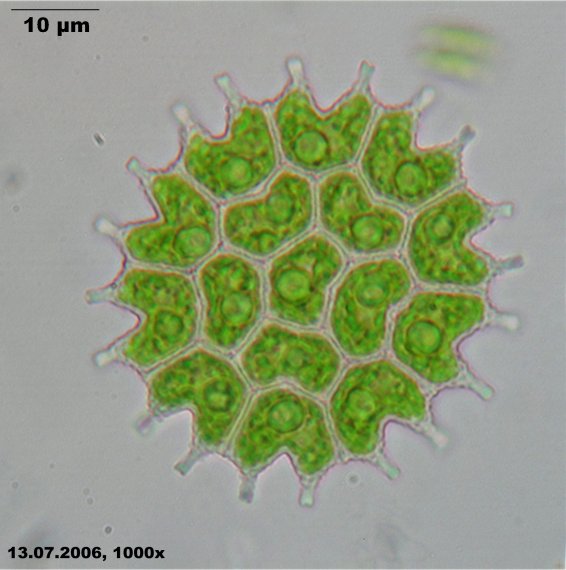
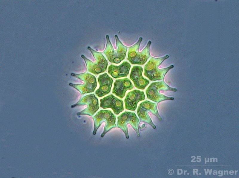
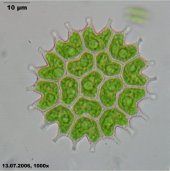
pediastrum boryanum
garden pond,
Bieberbach
July 2006 |
10 pictures stacked with CombineZ5 |
|
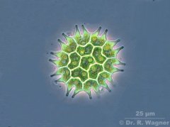
Pediastrum boryanum
Fishpond near Denkligen,
October 2007 |
Phasecontrast, 3 pictures stacked with CombineZM |
|
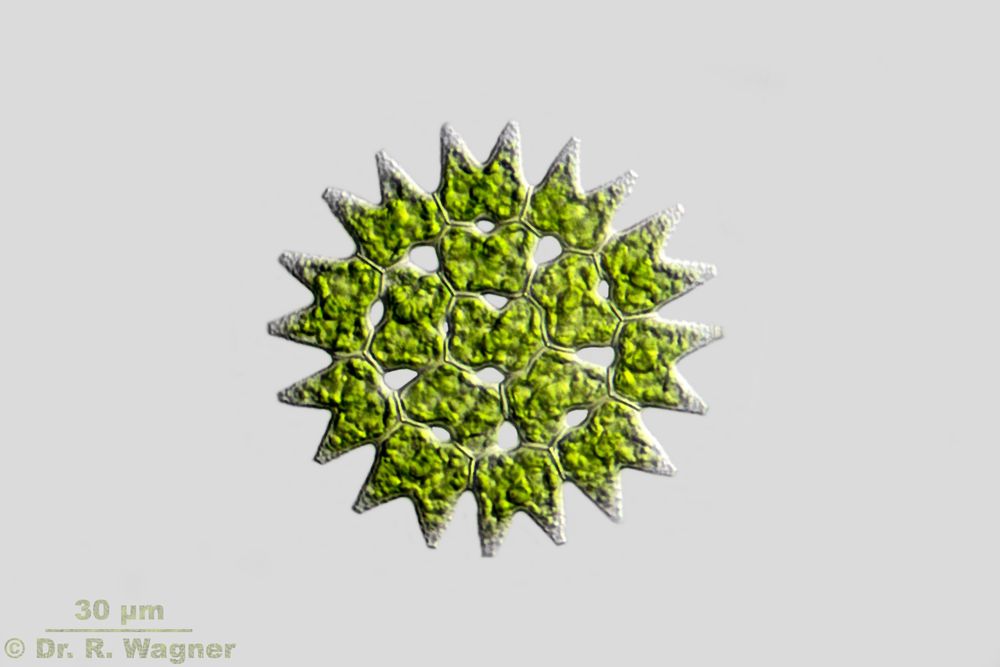
pediastrum duplex
botanical garden university Duesseldorf,
August 2003 |
|
 |
Pediastrum duplex var. subgranulatum
gardenpond
August 2016 |
|

|
pediastrum gracillimum
little creek near Roermond
October 2002 |
|
|
pediastrum tetras
botanical garden university Duesseldorf,
December 2006 |
|

|
scenedesmus spec.
garden pond,
spring 2001 |
|

|
scenedesmus acuminatus
garden pond,
spring 2001 |
|
|
Scenedesmus disciformis
Southpark, Düsseldorf
August 2008 |
|

|
scenedesmus longispina
botanical garden university Duesseldorf,
May 2005 |
|

|
scenedesmus opoliensis
garden pond,
spring 2001 |
|
 |
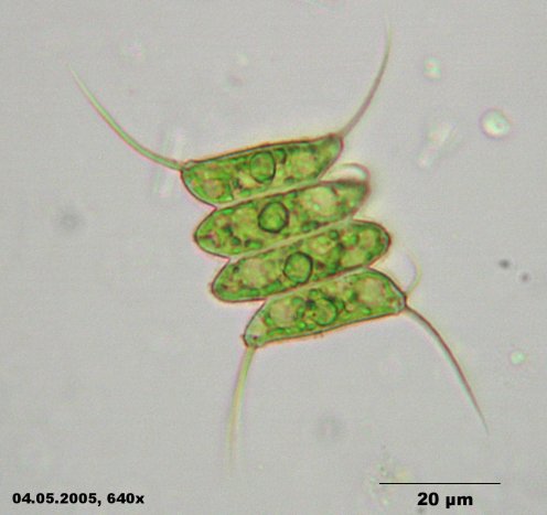
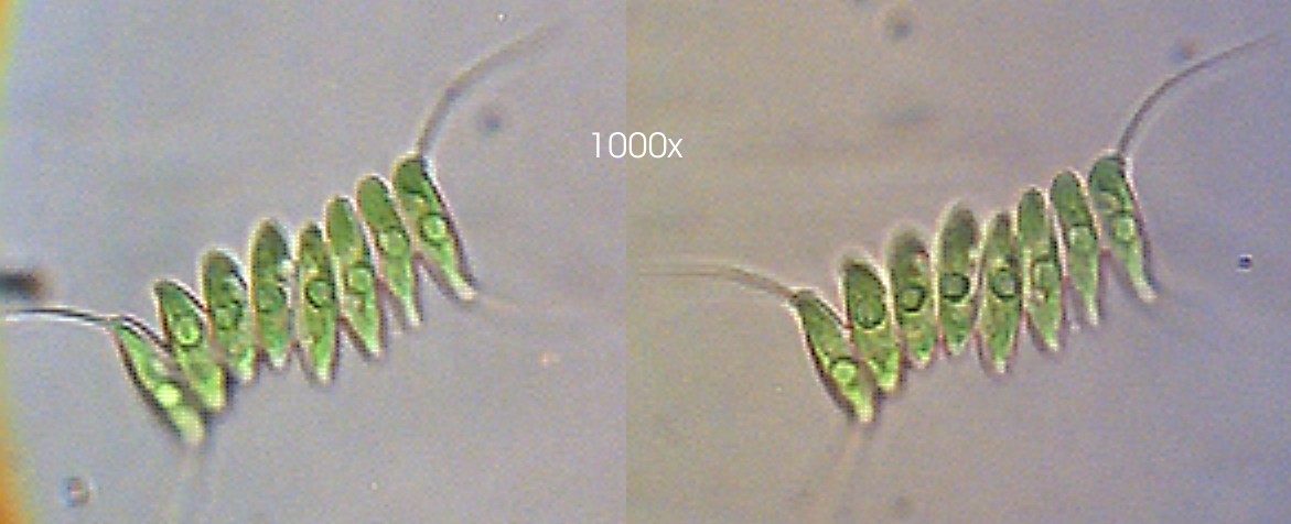
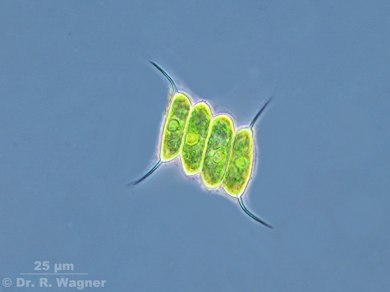
Scenedesmus quadricauda
Elfenmeer, a heathland pond,
August 2008 |
Phasecontrast |
 |
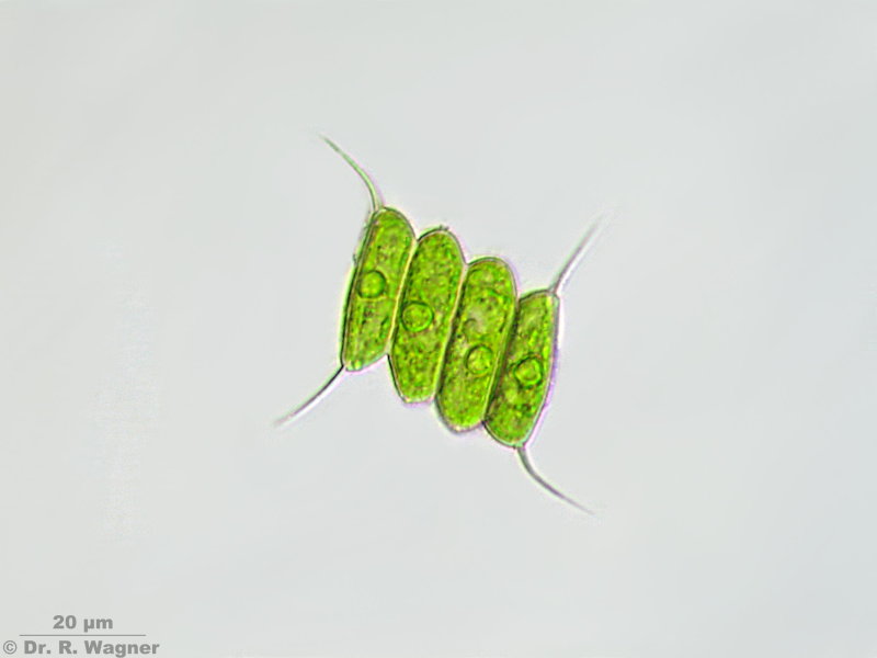
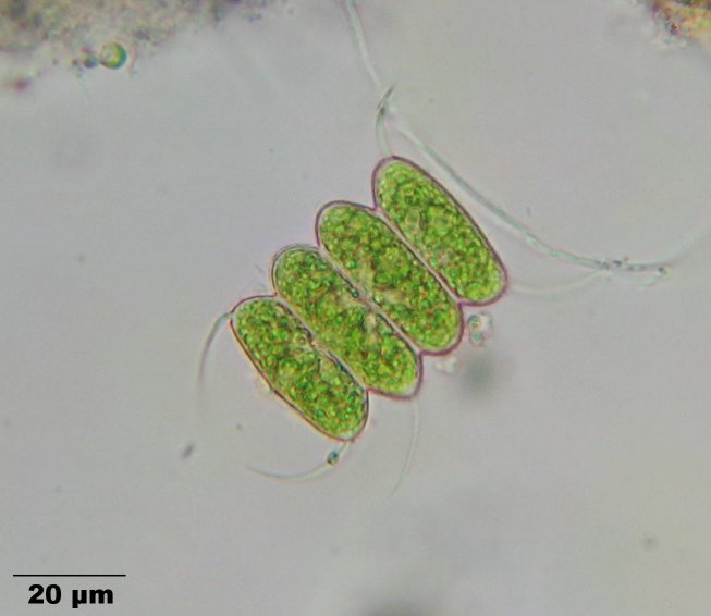
Scenedesmus quadricauda
Elfenmeer, a heathland pond,
August 2008 |
|
|
scenedesmus tenuispina
botanical garden university Duesseldorf,
March 2006
|
5 pictures stacked with CombineZ5. |
|
selenastrum bibraianum
national horticultural show Duesseldorf,
September 2006
|
|
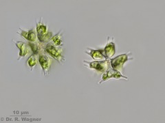 |
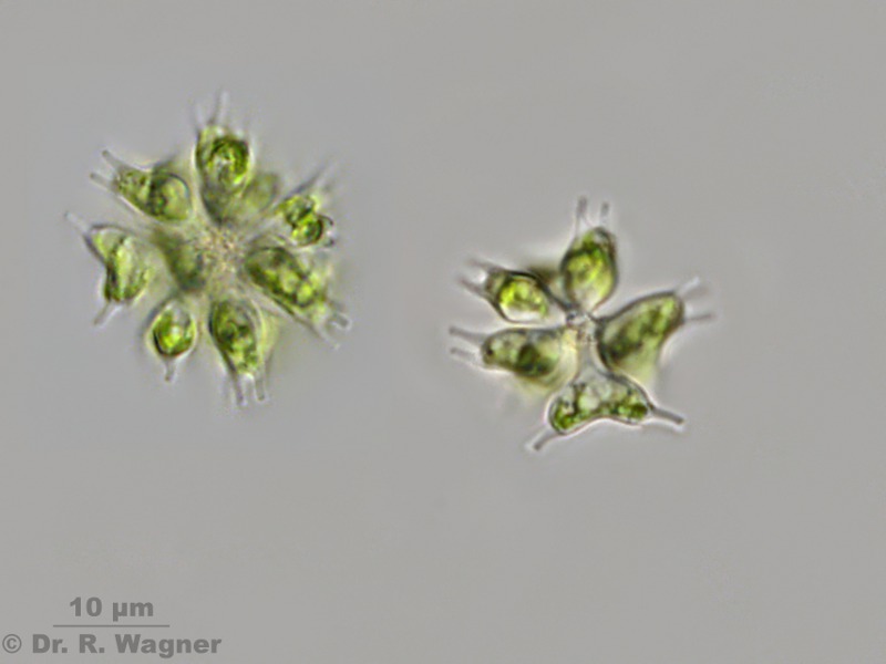
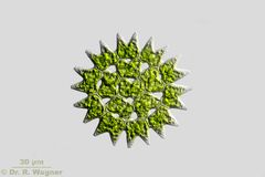
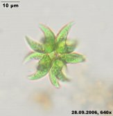
Sorastrum spinulosum
Small pond near Hilden,
May 2011 |
|

|
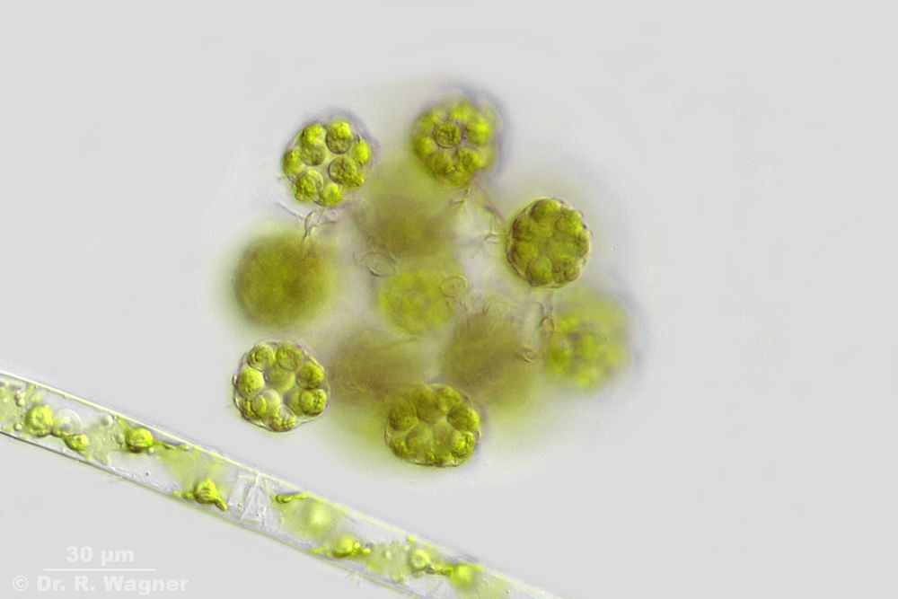
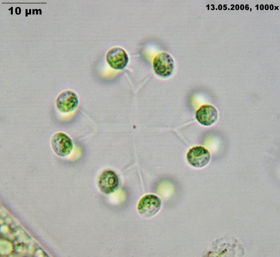
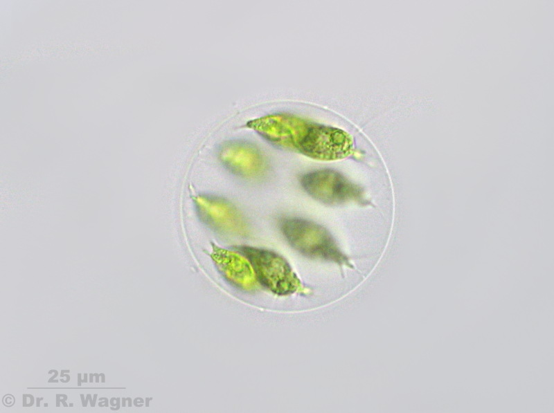
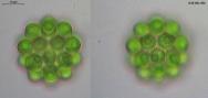
Stephanosphaera pluvialis
Bird bath, Müden
July 2008 |
Stephanospaera is related to Haematococcus. 8 cells form a ring in a common gelatin.
|

|
Stephanosphaera pluvialis
Bird bath, Müden
July 2008 |
Each cell has two flagella, an eyespot, several contractile vacuoles and a large chloroplast with several pyrenoids.
|
|
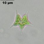
tetraedron caudatum
botanical garden university Duesseldorf,
September 2006
|
|

|
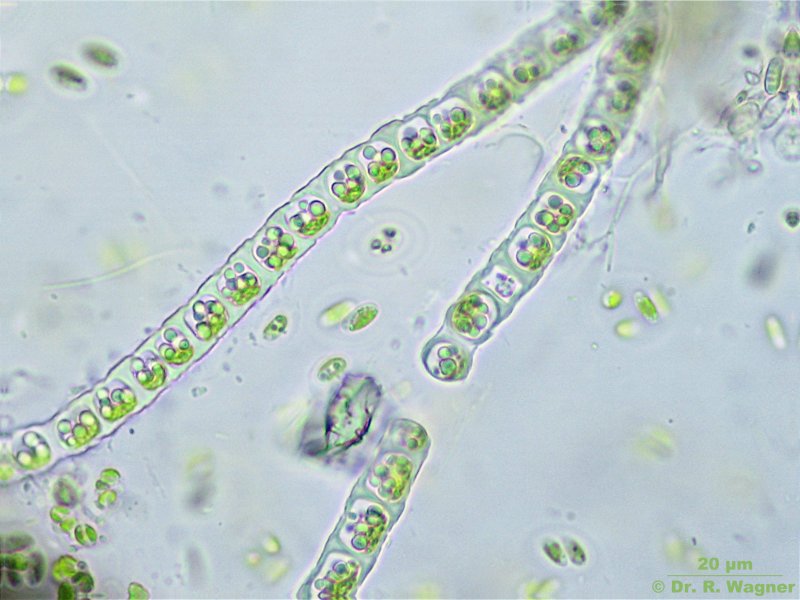
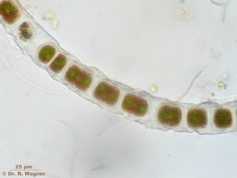
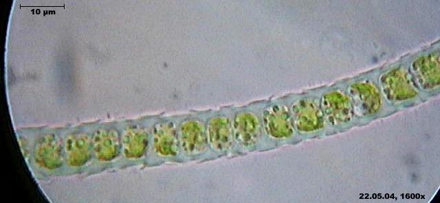
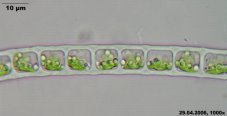
ulothrix moniliformis
Cologne-Wahn moor
May 2004
|
|

|
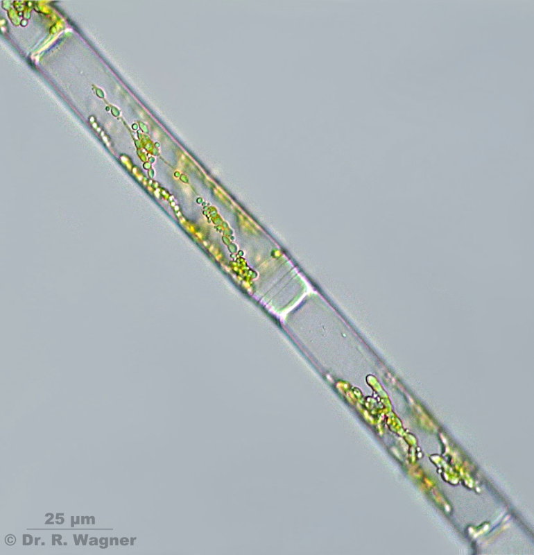
vaucheria spec. mit synplanospore
Itter-creek, Hilden
March 2003
|
|
 |
|
This links opens a seperate page on the theme
VOLVOX |