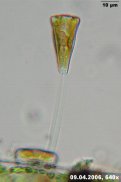
| preview-picture |
description |
comment |
|
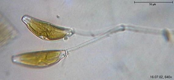
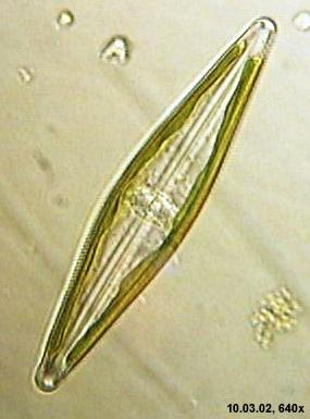
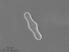
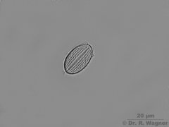
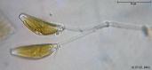
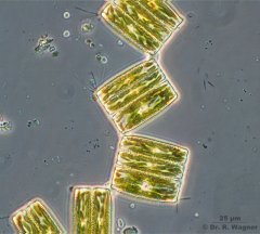
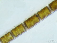
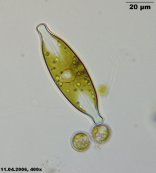
achnanthes clevel
Unterbacher pond, Duesseldorf
July 2002 |
girdle viewMicroscopy, photomicrograph, Microfoto |

|
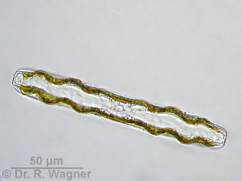
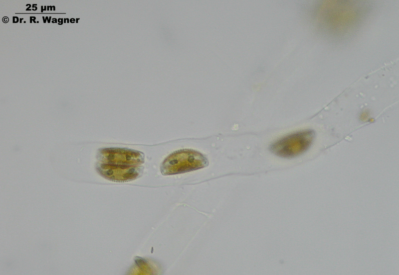
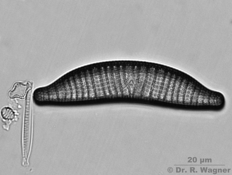
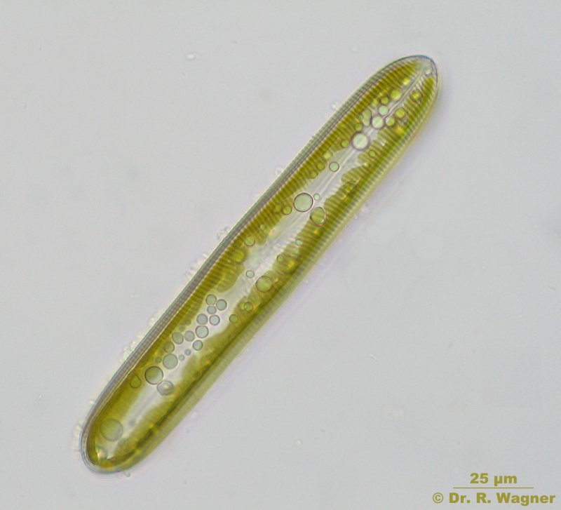
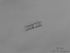
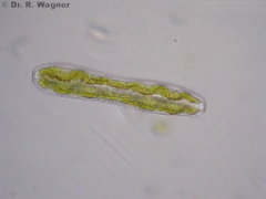
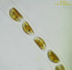
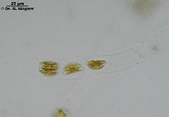
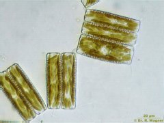
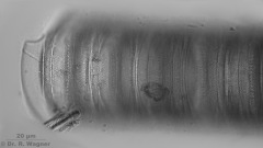
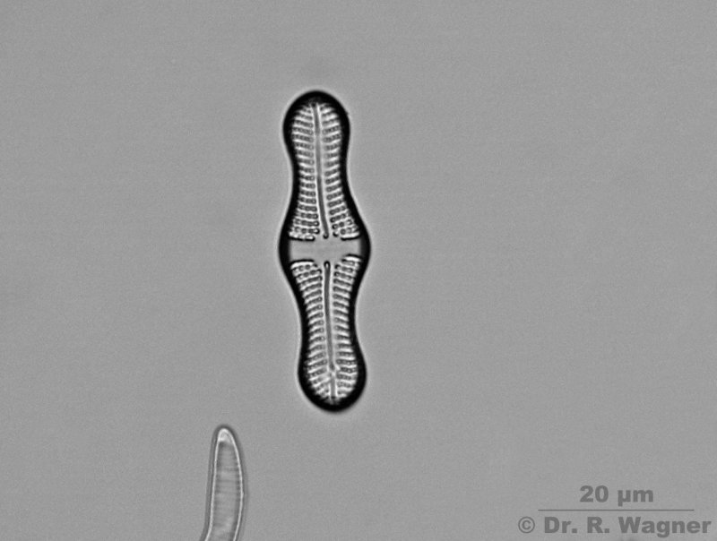
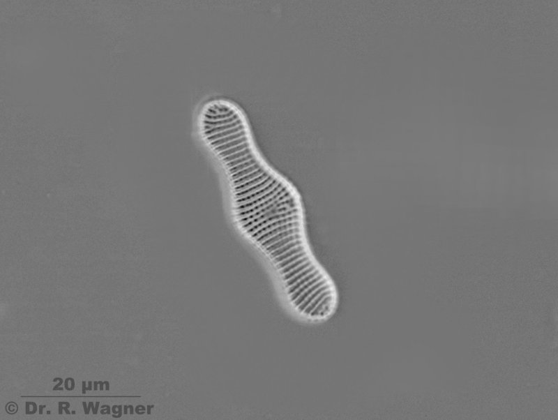
Achnanthes inflata
Aquarium
January 2008 |
Girdle view of the living cellMicroscopy, photomicrograph, Microfoto |
|
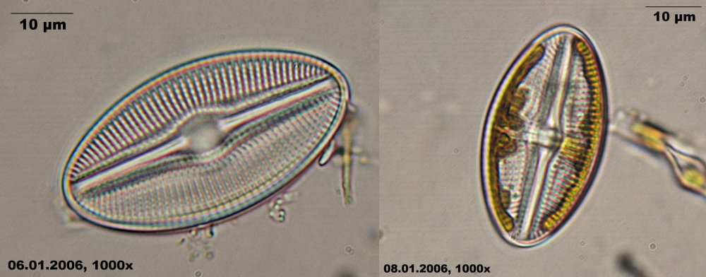
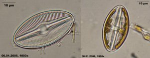
Achnanthes inflata
Aquarium
January 2008 |
RapheMicroscopy, photomicrograph, Microfoto |
|
Achnanthes inflata
Aquarium
January 2008 |
Side without rapheMicroscopy, photomicrograph, Microfoto |
 |
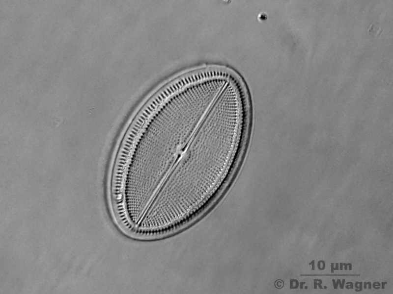
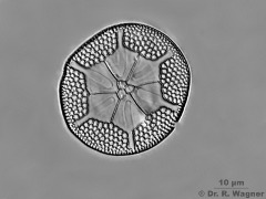
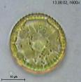
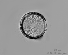
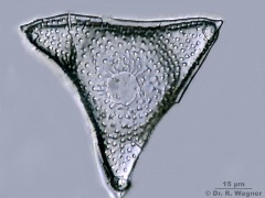
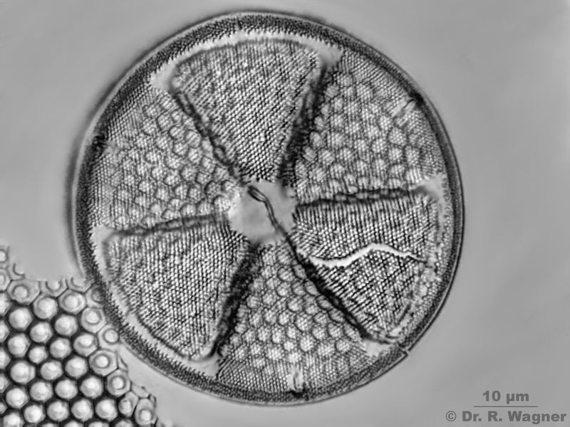
Actinoptychus senarius Ehrenberg
Desbarrancado
Peru |
Diameter 63 µmMicroscopy, photomicrograph, Microfoto |
|
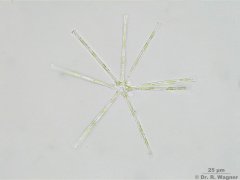
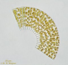
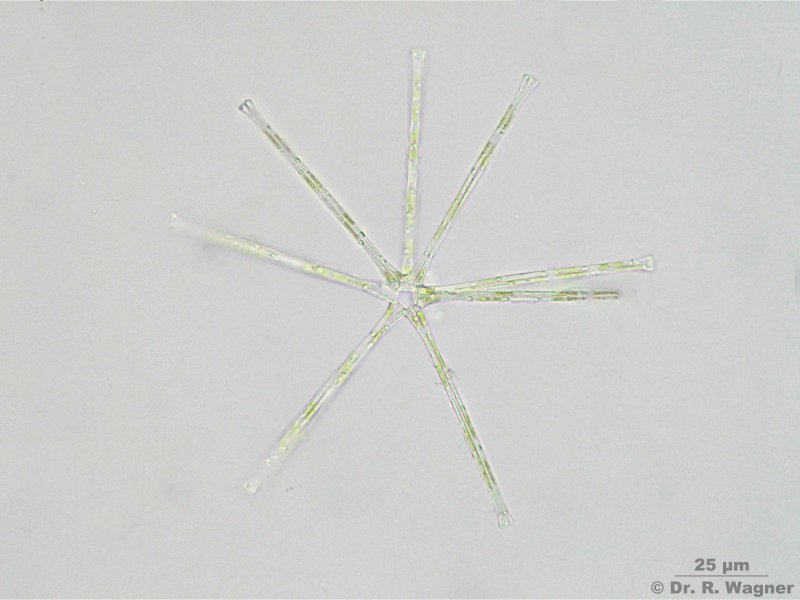
Asterionella formosa
quarry pond near Hilden
August 2007 |
Microscopy, photomicrograph, Microfoto |
|
 |
Asterolampra affinis
Desbarrancado
Peru |
Microscopy, photomicrograph, Microfoto |
|
Aulacoseira spec.
Diatomite
Lüneburger Heide
Fossil (350000 years) |
ZraxMicroscopy, photomicrograph, Microfoto |
|
-07_K.jpg)
|
Bacillaria paxillifer (former B. paradoxa)
Ruhr near Witten
July 2007 |
The cells of B. paxillifer tend to form clusters with each other. In these clusters the cells move along with
their raphes faced to their neighbours. The velocity of one cells is hereby added to that of the neighbour-cells and so
interesting movements occur. |
|
_8392_K.jpg)
|
Bacillaria paxillifer (former B. paradoxa)
Ruhr near Witten
July 2007 |
Video (divx-coded). The video shows the above described characteristic movement of B. paxillifer.Microscopy, photomicrograph, Microfoto |
|
_8400_K.jpg)
|
Bacillaria paxillifer (former B. paradoxa)
Ruhr near Witten
July 2007 |
Video (divx-coded). The video shows the above described characteristic movement of B. paxillifer. |
|
 |
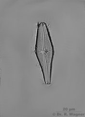
Cocconeis placentula
Botanical Garden,
University Duesseldorf
February 2008 |
Back side without raphe, 33 µm long, 22 µm wide, 18,5 stripes on 10 µm,
23 points on 10 µm
Microscopy, photomicrograph, Microfoto |
 |
Cocconeis placentula
Botanical Garden,
University Duesseldorf
February 2008 |
Front side with raphe, 33 µm long, 22 µm wide, 18,5 stripes on 10 µm,
23 points on 10 µm
Microscopy, photomicrograph, Microfoto |
 |
cyclotella spec.
little creek near Langenfeld
September 2005
|
Microscopy, photomicrograph, Microfoto |
 |
cyclotella comta
little creek near Roermond
October 2002
|
Microscopy, photomicrograph, Microfoto |
 |
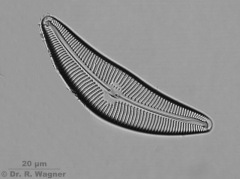
Cymatopleura solea
Southpark, Düsseldorf
March 2009 |
valve viewMicroscopy, photomicrograph, Microfoto |
 |
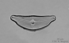
Cymatopleura solea
Southpark, Düsseldorf
March 2009 |
Girdle viewMicroscopy, photomicrograph, Microfoto |
 |
Cymatopleura solea
Southpark, Düsseldorf
March 2009 |
Video, 9 MBMicroscopy, photomicrograph, Microfoto |
 |
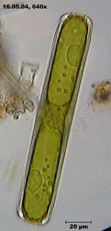
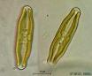
Cymbella cistula
Botanical Garden,
University Duesseldorf
February 2008 |
56 µm long, 13 µm wide, 10 striepes on 10 µm, 22 points on 10 µm
Microscopy, photomicrograph, Microfoto |

|
Cymbella helvetica
Diatomite
Lüneburger Heide
Fossil (350000 years) |
ZraxMicroscopy, photomicrograph, Microfoto |

|
cymbella lanceolata
garden pond
October 2003 |
Microscopy, photomicrograph, Microfoto |
|
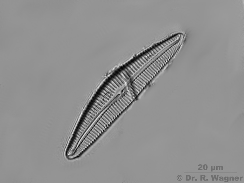
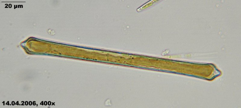
Cymbella proxima
botanical garden university Duesseldorf,
February 2008 |
lenght: 78 µm; width: 20 µm; 8 stripes per 10 µm; 14 pores per 10 µm
Microscopy, photomicrograph, Microfoto |
|
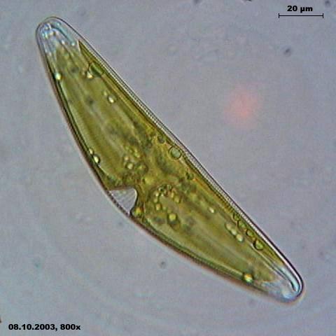
Cymbella tumida
botanical garden university Duesseldorf
April 2008 |
Zrax, length: 56 µm; width: 16 µm; 10 stripes per 10 µm; 18 pores /10 µm
Microscopy, photomicrograph, Microfot |
|
cymbella ventricosa
botanical garden university Duesseldorf,
July 2002 |
Microscopy, photomicrograph, Microfoto |
|
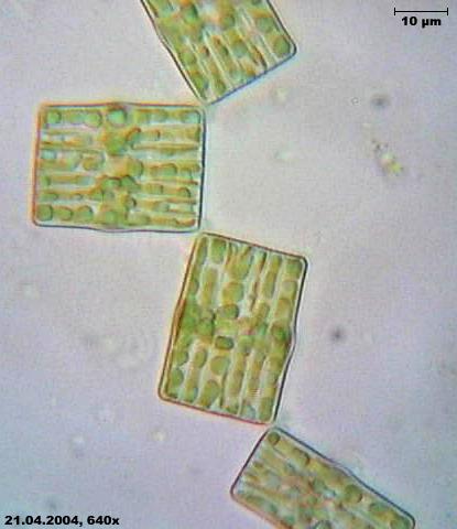
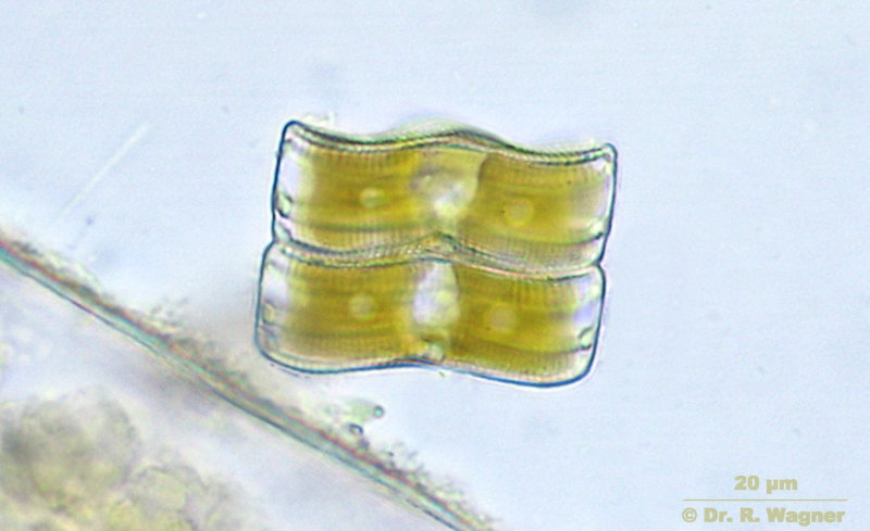
Cymbella spec.
South-Park,
Duesseldorf,
March 2007 |
Several algae in one common mucous borderline |
|
Cymbella spec.
South-Park,
Duesseldorf,
March 2007 |
Several algae in one common mucous borderline, in which they can move forwardMicroscopy, photomicrograph, Microfoto |
|
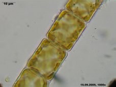
Diatoma vulgaris
Brook near Langenfeld
June 2007 |
Microscopy, photomicrograph, Microfoto |
|
Diatoma vulgaris
Brook near Langenfeld
June 2007 |
Microscopy, photomicrograph, Microfoto |
|
diploneis species
Schee tunnel, Wuppertal
January 2006 |
valve view, raphe good visible. the left picture shows only the skeleton
, the right picture a living individual. (CombineZ5, 10 pictures stacked)Microscopy, photomicrograph, Microfoto |
|
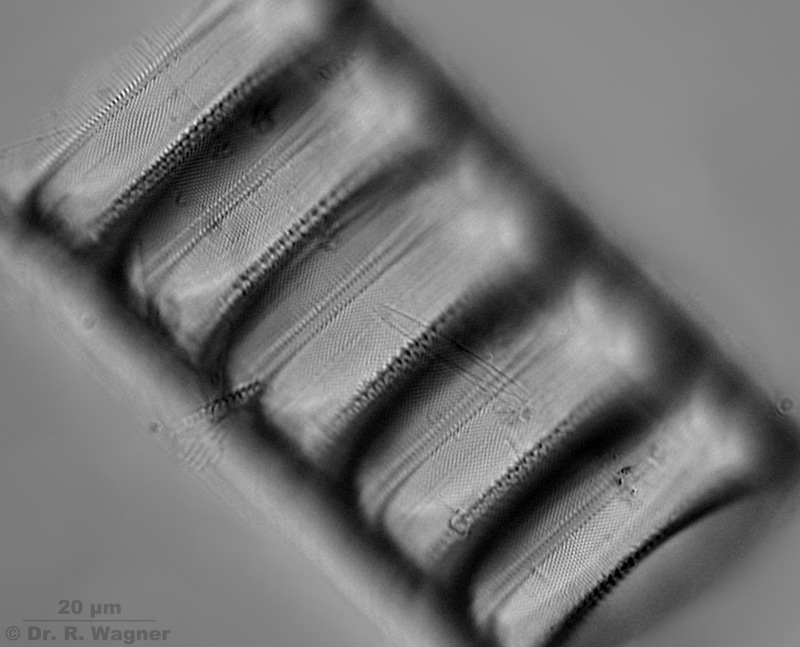
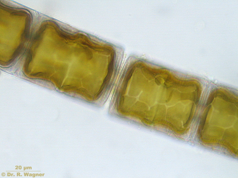
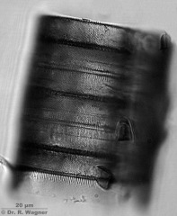
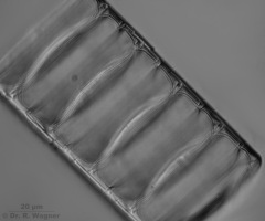
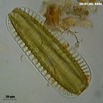
Ellerbeckia arenaria
Botanical garden
University Düsseldorf
April 2008 |
Similar to Melosira Ellerbeckia arenaria forms long chains. Mountant: airMicroscopy,
photomicrograph, Microfoto |
|
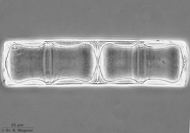
Ellerbeckia arenaria
Botanical garden
University Düsseldorf
April 2008 |
In addition to the main girdle band in the middle of each cell intermediate girdlebands
can be seen, too. Mountant: ZRAXMicroscopy, photomicrograph, Microfoto |

|
Ellerbeckia arenaria
Botanical garden
University Düsseldorf
April 2008 |
With the mountant Euparal contrasts are weaker, but on the other hand there is little more depth of focus.
Microscopy, photomicrograph, Microfoto |
|
Ellerbeckia arenaria
Botanical garden
University Düsseldorf
April 2008 |
Only with the mountant Euparal it is possible to show the surface line in an optical cross-section.
The contrats with ZRAX are too heavy and the picture gets too dark.Microscopy,
photomicrograph, Microfoto |
|
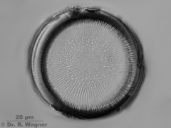
Ellerbeckia arenaria
Botanical garden
University Düsseldorf
April 2008 |
Discus is flt with radial stripes that do not reach the centre. Mountant: ZRAX
Microscopy, photomicrograph, Microfoto |
|
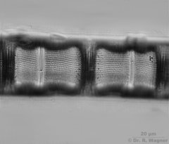
Epithemia adnata
Diamtomite
Lüneburger Heide
Fossil (350000 years) |
ZraxMicroscopy, photomicrograph, Microfoto |
|
Epithemia sorex
Diamtomite
Lüneburger Heide
Fossil (350000 years) |
ZraxMicroscopy, photomicrograph, Microfoto |

|
Epithemia turgida
Botanical garden
University Düsseldorf |
NaphraxMicroscopy, photomicrograph, Microfoto |

|
eunotia valida
Hilden heathland,
December 2006 |
Microscopy, photomicrograph, Microfoto |
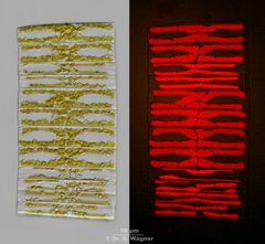 |
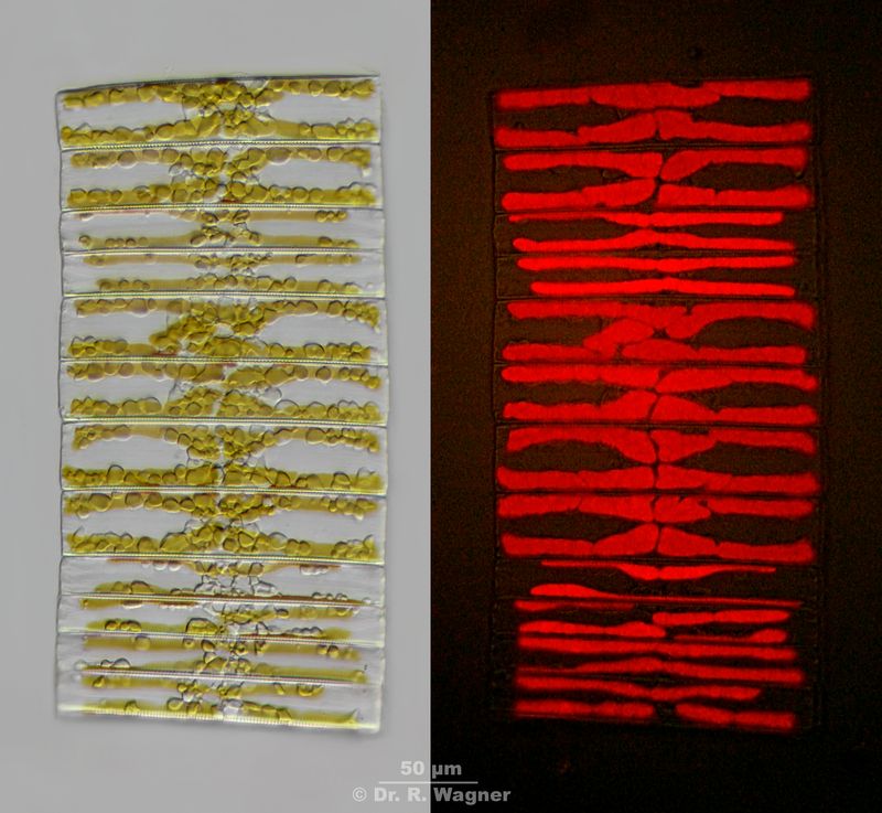
Fragilaria spec.
between Sphagnum,
Birgelen, virgin forest
April 2013 |
Brightfield and Chlorophyll autofluorescence in 365 nm UV-excitation
Microscopy, photomicrograph, Microfoto |
|
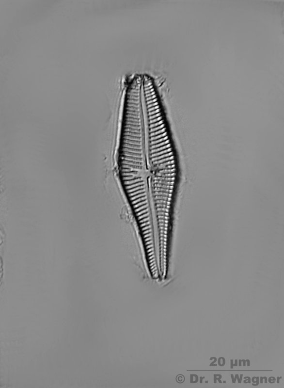
gomphonema spec.
national horticultural show Duesseldorf,
April 2006 |
10 pictures stacked with CombineZ5 |
|
Gomphonema ventricosum
Diamtomite
Lüneburger Heide
Fossil (350000 years) |
ZraxMicroscopy, photomicrograph, Microfoto |
|
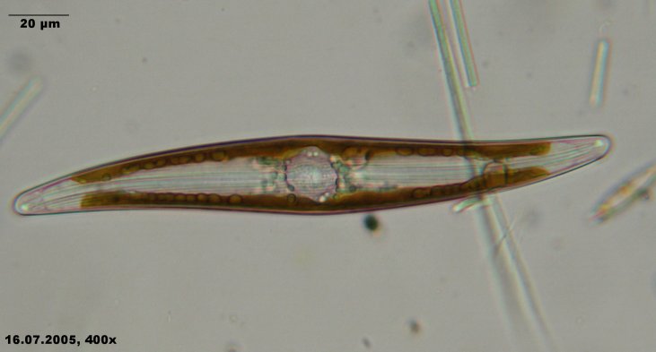
gyrosigma spec.
bathing pond in the Ville
July 2005 |
Microscopy, photomicrograph, Microfoto |
 |
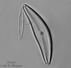
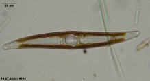
Gyrosigma acuminatum
Botanical garden,
University Düsseldorf,
April 2008 |
Stripes 13 / 10 µm, pores 12 / 10 µm). Size: 125 µm long, 19 µm broad.
Microscopy, photomicrograph, Microfoto |
|
Melosira undulata
freshwater aquarium
January 2007 |
Microscopy, photomicrograph, Microfoto |

|
Melosira undulata
freshwater aquarium
January 2007 |
Microscopy, photomicrograph, Microfoto |

|
Melosira undulata
freshwater aquarium
January 2007 |
annealed preparation, phase contrastMicroscopy, photomicrograph, Microfoto |
|
Melosira undulata
freshwater aquarium
January 2007 |
annealed preparation, approx. 16 rows of pores pro 10 µm along the longitudinal axis.
There are wart-like dots on the left and right cell ends.Microscopy, photomicrograph, Microfoto |
|
Melosira undulata
freshwater aquarium
January 20077 |
annealed preparation. The discus shows radial, sometimes branched, rows of pores,
that let the center free.Microscopy, photomicrograph, Microfoto |
|
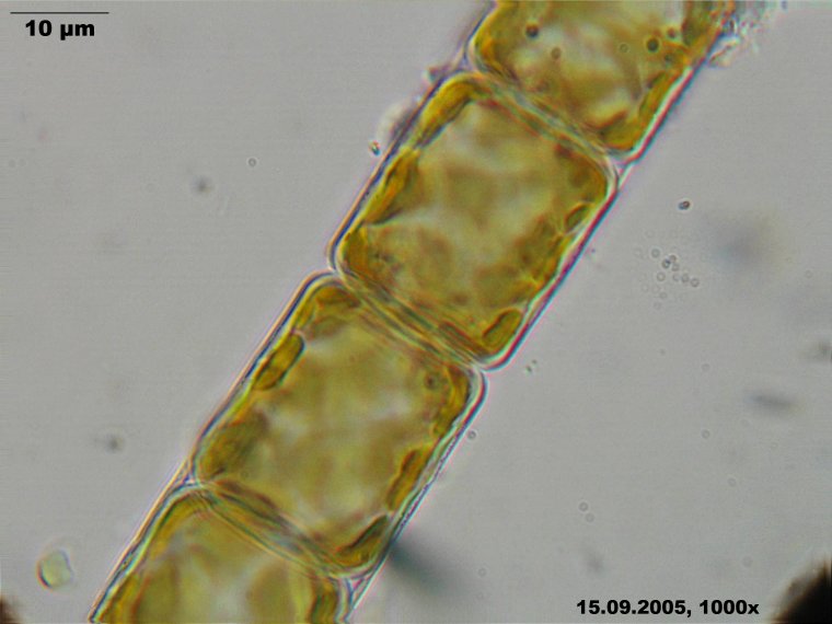
melosira varians
little creek near Langenfeld Uferzone
September 2005 |
CombineZ5, 3 pictures |

|
melosira varians
Urdenbacher floodplain,
May 2004 |
formation of an auxozygoteMicroscopy, photomicrograph, Microfoto |
|
Meridion circulare
Schee tunnel,
April 2007 |
Microscopy, photomicrograph, Microfoto |
 |
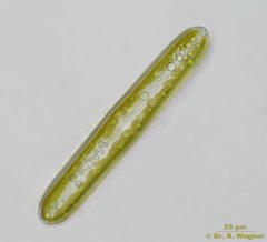
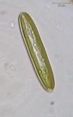
navicula radiosa
Hilden heathland, March 2002 |
Microscopy, photomicrograph, Microfoto |
|
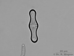
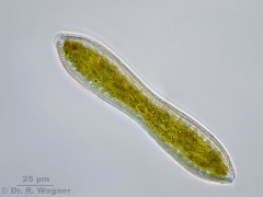
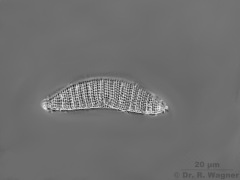
pinnularia spec,
Schwalm-Nette area, white stone,
December 2006 |
valve view |
|
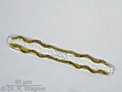
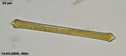
pinnularia viridis
Urdenbacher floodplain
May 2004 |
GürtelbandansichtMicroscopy, photomicrograph, Microfoto |
|
pinnularia_viridis
garden pond
December 2002 |
video (0,62 MB, avi-format)Microscopy, photomicrograph, Microfoto |
|
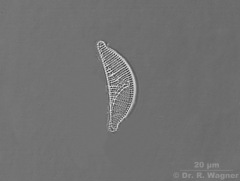
stauroneis anceps
national horticultural show Duesseldorf,
April 2006 |
5 pictures stacked with CombineZ5 |
|
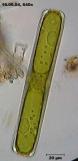
stauroneis smithii
garden pond
August 2002 |
Microscopy, photomicrograph, Microfoto |
|
surirella robusta splendida
Cologne-Wahn moor
July 2004 |
Microscopy, photomicrograph, Microfoto |
|
synedra capitata
botanical garden university Duesseldorf,
April 2006 |
Microscopy, photomicrograph, Microfoto |
|
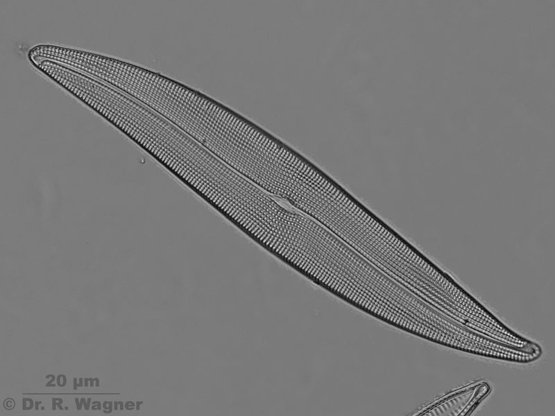
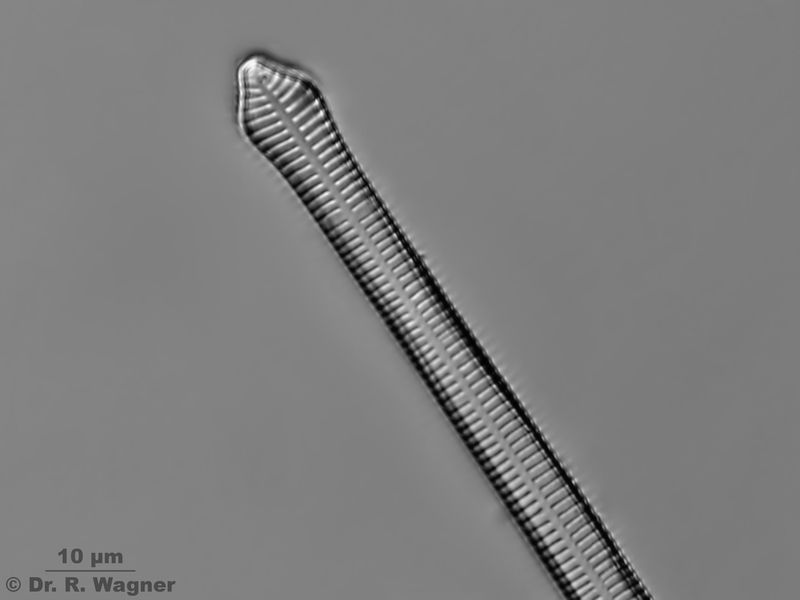
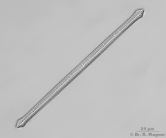
Synedra capitata
botanical garden university Duesseldorf,
February 2008 |
Naphrax, Sulfuric acid, Potassiumpermanganate, Oxalic acid
Length: 190 µm, Width: 8 µm, 11 Stipes / 10 µmMicroscopy, photomicrograph, Microfoto |

|
Synedra capitata
botanical garden university Duesseldorf,
February 2008 |
Naphrax, Sulfuric acid, Potassiumpermanganate, Oxalic acid
Length: 190 µm, Width: 8 µm, 11 Stipes / 10 µmMicroscopy, photomicrograph, Microfoto |
 |
tabellaria flocculosa
creek near Obersteinbach (F), Alsace
April 2004 |
Microscopy, photomicrograph, Microfoto |
 |
Trinacria ventricosa
Desbarrancado
Peru |
Microscopy, photomicrograph, Microfoto |


-07.jpg)