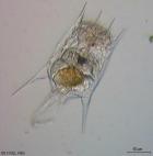
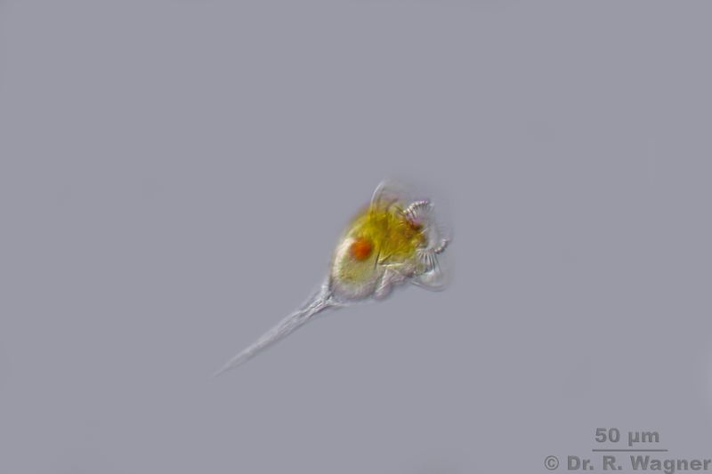
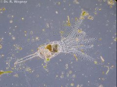
Conochilus unicornis Quarry pond near Hilden, March 2014
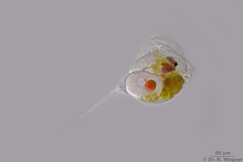
Conochilus unicornis Quarry pond near Hilden, July 2016
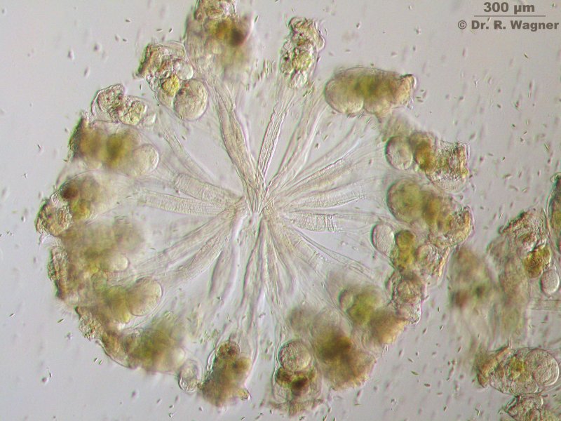
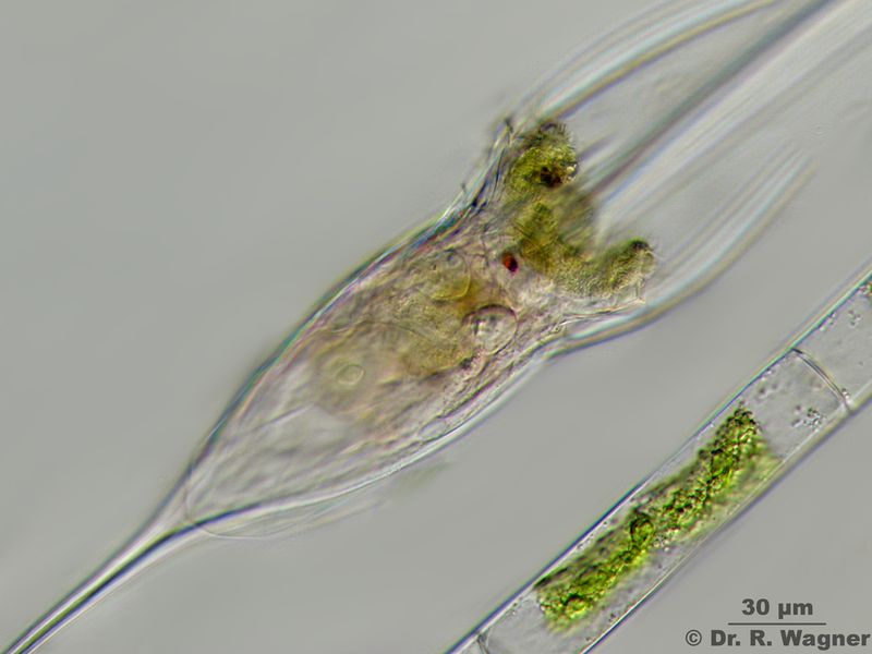
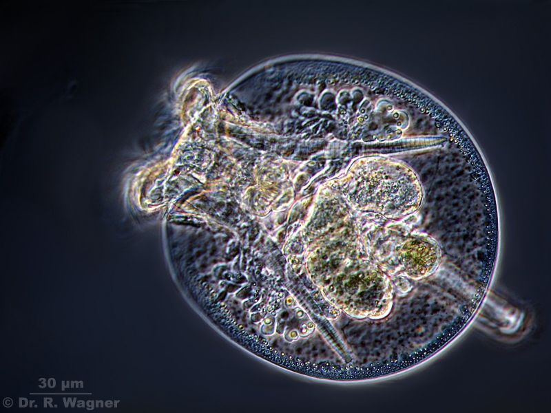
Conochilus unicornis Quarry pond near Hilden, August 2007Microscopy, photomicrograph, Microfoto
Conochilus unicornis forms ball shaped colonies with up to 25 individuals, each in its own gelantine.
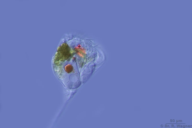
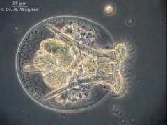
Conochilus unicornis Quarry pond near Hilden, August 2007
Microscopy, photomicrograph, Microfoto
Conochilus unicornis Quarry pond near Hilden, August 2007
Microscopy, photomicrograph, Microfoto
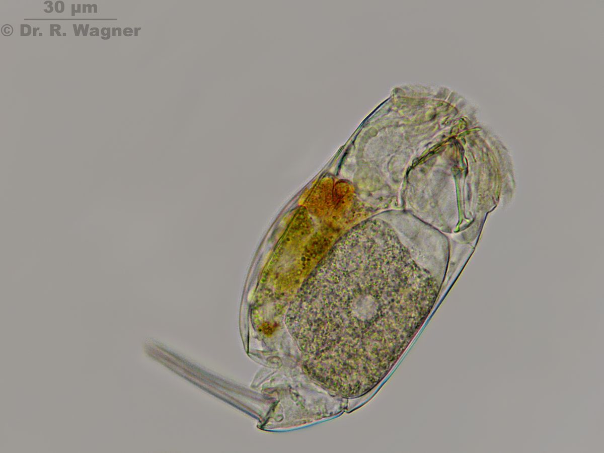
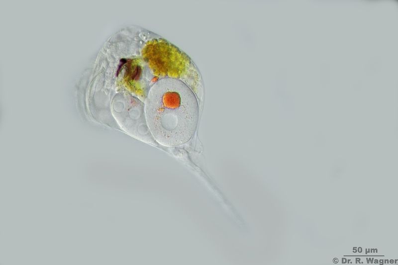
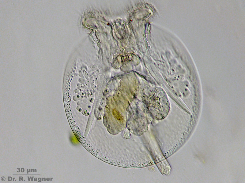
Conochilus unicornis Quarry pond near Hilden, August 2007Microscopy, photomicrograph, Microfoto
Female with an egg inside. Inside the egg you can see parts of the mastax and the red eye of the hatchling.
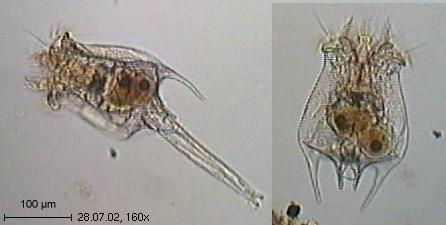
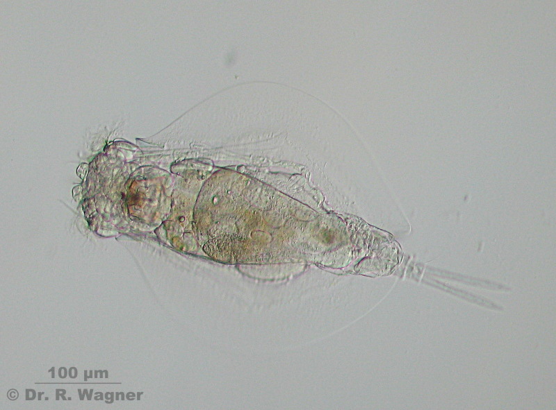
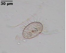
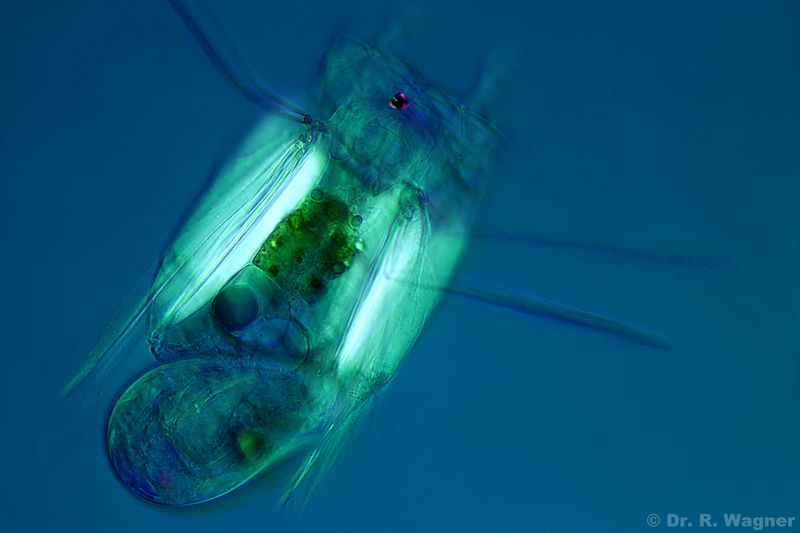
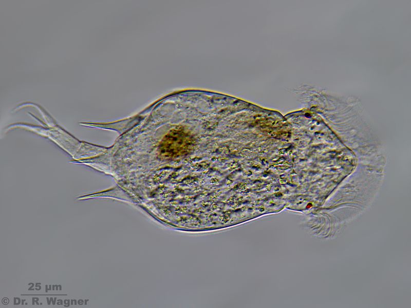
Euchlanis triquetra Southpark, Düsseldorf, November 2007Microscopy, photomicrograph, Microfoto
Oblique illumination. The carapace is very transparant. It can be seen with oblique illum. or in phase contrast, only.
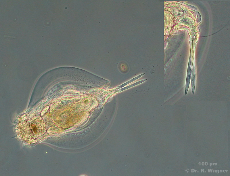
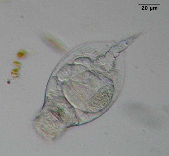
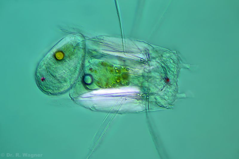
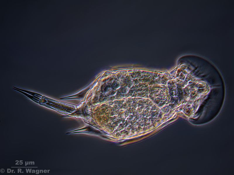
Euchlanis triquetra Southpark, Düsseldorf, November 2007Microscopy, photomicrograph, Microfoto
Phasecontrast. The inlet shows one of the two characteristic bristles at the foot.
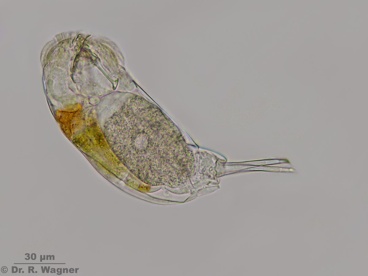
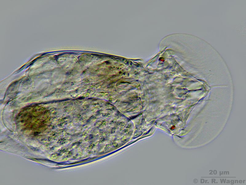
Euchlanis triquetra Southpark, Düsseldorf, November 2007
A close view of the head with the eyespot and the mastax.Microscopy, photomicrograph, Microfoto
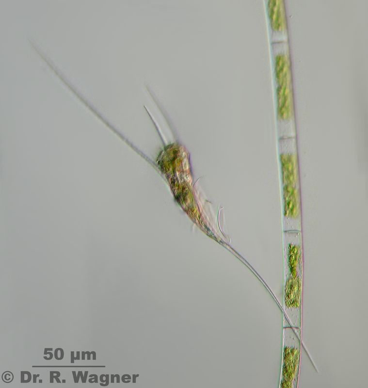
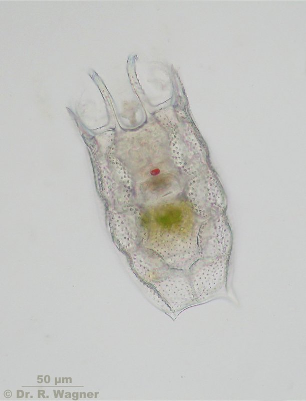
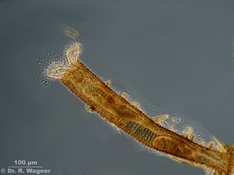
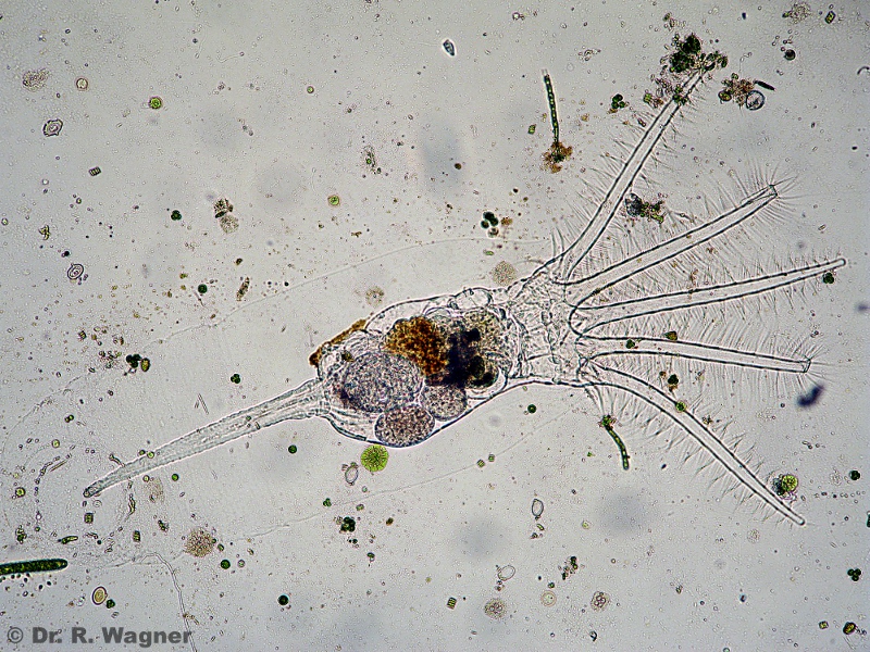
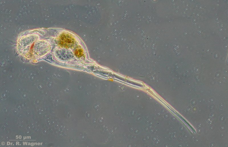
Kellicottia longispina Quarry near Hilden, April 2011
Front border with long spines. Back with one one very lomng spine. Planktic.
Microscopy, photomicrograph, Microfoto
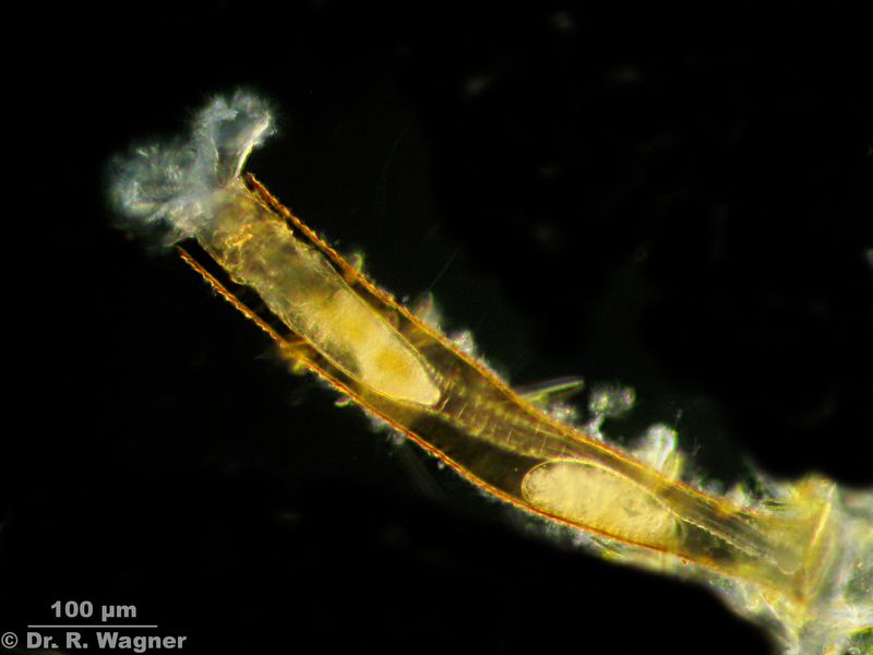
Kellicottia longispina Quarry near Hilden, April 2011
Closeup with eyespotMicroscopy, photomicrograph, Microfoto
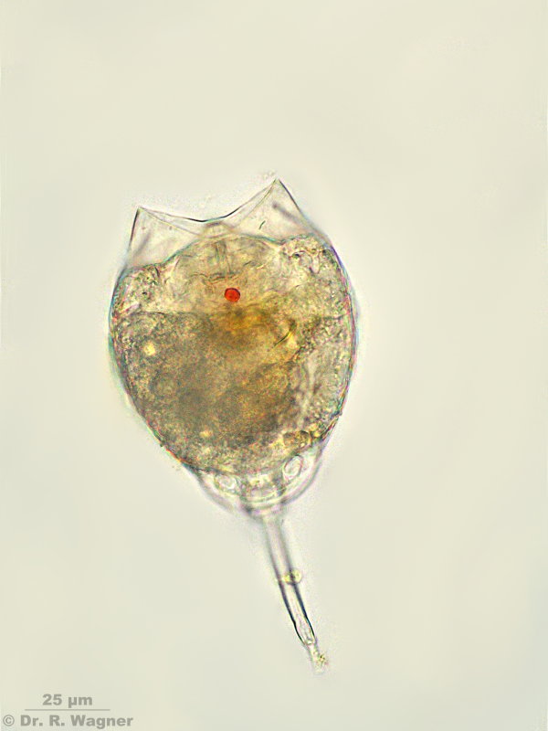
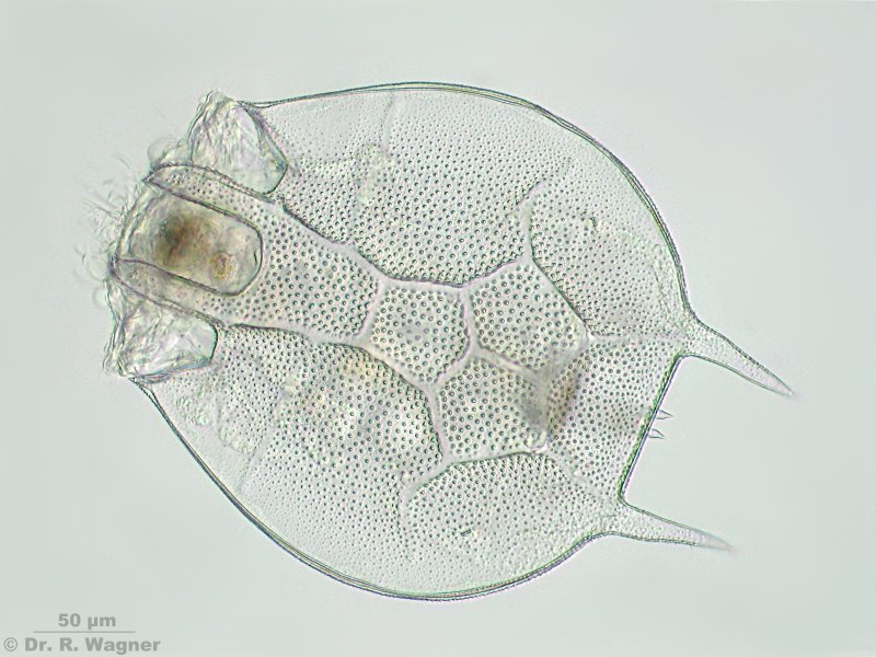
Keratella quadrata Weisser Stein, heathland December 2006
Microscopy, photomicrograph, Microfoto
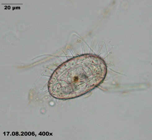
Lecane lunaris Pietz-Moor, Lüneburger Heide July 2008
Microscopy, photomicrograph, Microfoto
lepadella patella botanical garden university Duesseldorf, August 2006
lepadella patella in its eggMicroscopy, photomicrograph, Microfoto
lepadella patella botanical garden university Duesseldorf, August 2006
video, divx-format. In the lower half of the picture you can see the foot and in the middle the mastax.
The mastax is already working, as you can see in the videoMicroscopy, photomicrograph, Microfoto
lepadella patella botanical garden university Duesseldorf, August 2006